(We are very thankful that Yngve Kristiansen, PhD candidate at the University of Bergen VisGroup could write this report looking forward to the medical visualization-related papers at EuroVis 2022 for us!)
It is now the time, EuroVis is coming up and there are a lot of new exciting papers. If you are interested in medical visualization, there are many papers for you! But they are spread across different sessions, so I thought it would be nice to create a schedule for medical-relevant contributions at this year’s EuroVis! Preprints and information is available for most of the papers, but some of them were not yet published, but I still decided to put them in the schedule because I think they are definitely worth checking out!
Monday
(Paper) Toward disease diagnosis visual support bridging classic and precision medicine
Alessia Palleschi, Manuela Petti, Paolo Tieri, and Marco Angelini
Monday June 13th, EuroVA 3 : Applications – Aula Magna Room @ 14:20 – 14:45
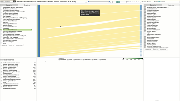
The authors present a visual analytics approach for helping medical personnel investigate symptoms, anatomies, diseases and genes during the diagnosis process. The goal of their tool is to combine the classic, often symptom-based appraoch with precision medicine — which considers more factors such as interactions between diseases, individual genes and other factors. They provide a visual analysis environment where the clinician sees a sankey diagram to compare similarities across multiple diseases.
(Paper) Physical Traces and Digital Stories: Exploring the Connections Between Forensics and Visualization
Victor Schetinger and Saminu Salisu
Monday June 13th, VisGap 1 : Domain Considerations – Sala Colonne Room A @ 15:35 – 15:55
No more information or preprint available at the time of writing.
Tuesday
(Poster) Automatic segmentation of tooth images: Optimization of multi-parameter image processing workflow
Giovani Bressan Fogalli, Daniel Baum, and Sergio Line
Tuesday June 14th, Welcome & Poster – Angelicum Garden @ 19:00
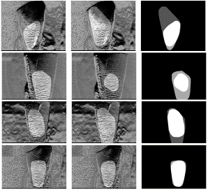
The authors propose an approach for automatically segmenting out Hunter-Schreger Bands from images of laterally illuminated teeth. They do this in the form of an image processing pipeline called anisotropy-based segmentation which they describe more closely in their poster.
(Poster) Explorative Visual Analysis of Spatio-temporal Regions to Detect Hemodynamic Biomarker Candidates
Adrian Derstroff, Simon Leistikow, Ali Nahardani, Mahyasadat Ebrahimi, Verena Hoerr, and Lars Linsen
Tuesday June 14th, Welcome & Poster – Angelicum Garden @ 19:00
No more information or preprint available at the time of writing.
(Poster) Visual Exploration of Genetic Sequence Variants in Pangenomes
Astrid van den Brandt, Eef M. Jonkheer, Dirk-Jan M. van Workum, Sandra Smit, and Anna Vilanova
Tuesday June 14th, Welcome & Poster – Angelicum Garden @ 19:00
No more information or preprint available at the time of writing.
(Poster) Accurate molecular atom selection in VR
Elena Molina Lopez and Pere-Pau Vázquez
Tuesday June 14th, Welcome & Poster – Angelicum Garden @ 19:00
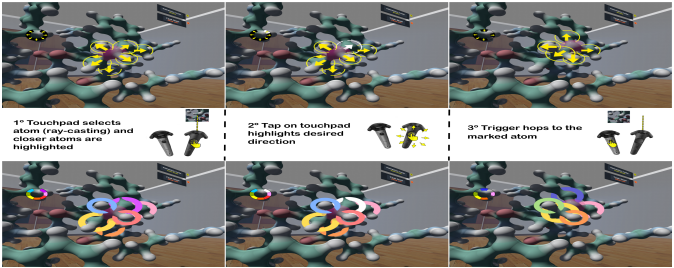
The authors present two new methods for selecting atoms in cluttered VR environments. Both methods are based on the same two following steps: (1) the user selects a single atom, (2) the system automatically highlights its neighbors, which the user can then select interactively.
Wednesday
(Paper) Barrio: Customizable Spatial Neighborhood Analysis and Comparison for Nanoscale Brain Structures
Jakob Troldi, Corrado Cali’, Eduard Gröller, Hanspeter Pfister, Markus Hadwiger, and Johanna Beyer
Wednesday June 15th, FP 5 : Life Sciences and Urbanism – Aula Minor Room @ 9:00 – 9:25

The authors present Barrio – a configurable framework for neuroscientists to explore brain structures at a nano scale. It is configurable to several scales, so that it can be configured to answer multiple tasks and domain questions. It provides a scalable approach for comparative visualization of such spatial neighborhood relationships. They do this by providing small multiples of spatial 3D views in addition to quantitative 2D views.
(STAR) Trends & Opportunities in Visualization for Physiology: A Multiscale Overview
Laura Garrison, Ivan Kolesar, Ivan Viola, Helwig Hauser, and Stefan Bruckner
Wednesday June 15th, STAR 2 : A Journey Through Multiple Scales – Aula Minor Room @ 15:10 – 16:00
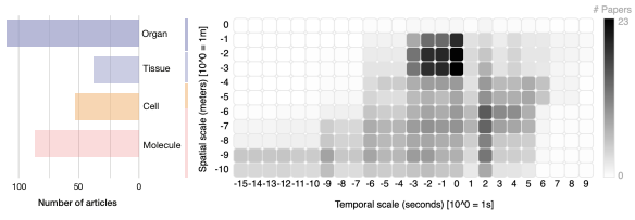
The authors summarizes trends and opportunities in visualization for physiology. They do this on two levels. On the first level, they sort literature according to organizational complexity, ranging from molecule to organ. Secondly, they classify which high-level visualization tasks (exploration, analysis, and communication) are used within an existing work.
Thursday
(STAR) Vessel Maps: A Survey of Map-Like Visualizations of the Cardiovascular System
Pepe Eulzer, Monique Meuschke, Gabriel Mistelbauer, and Kai Lawonn
Thursday June 18th, STAR 3 : Health and Medicine – Aula Magna Room @ 14:20 – 15:10
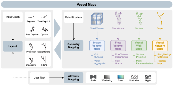
The authors make efforts to unify the vast amount of map-like visualizations of cardiovascular structures. They first provide a perspective of different data spaces from which map-like structures can be derived from. Then, they view existing approach in the light of a wide range of requirements from medical practitioners and researchers. Based on these perspectives, they offer recommendations for designing such map-like visualizations of the cardiovascular system.
(Paper) EHR STAR: The State-Of-the-Art in Interactive EHR Visualization
Qiru Wang and Robert S. Laramee
Thursday June 18th, STAR 3 : Health and Medicine – Aula Magna Room @ 15:10– 16:00
The authors provide the state-of-the-art for interactive electronic health record visualizations. They provide an up-to-date overview of terminology and recent research in the field of EHR. They survey 34 high quality open access healthcare data sources and datasets and 51 EHR visualization publications, and provide a classification with six re-occuring research themes and the Unified Medical Language System (UMLS).
(Paper) Nested Papercrafts for Anatomical and Biological Edutainment
Marwin Schindler, Thorsten Korpitsch, Renata Georgia Raidou, and Hsiang-Yun Wu
Thursday June 16th, FP 13 : General Public – Aula Minor Room @ 15:10-15:35
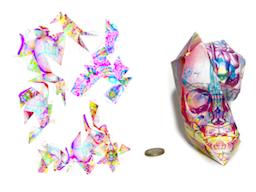
The authors present a new workflow for computer-aided generation of physicalizations, namely papercrafts, addressing nested configurations in anatomical and biological structures, seen from optimal viewpoints. Their approach aids the user by generating a paper figure version of an imported 3D model. This generated paper figure can then be printed and folded into a physicalization of the object.
Let us know if there are any papers or posters we missed in the comments!

Great that you prepared this. Thanks a lot!