Call for Participation: MedVis-Award 2016

The 7th “Karl-Heinz-Höhne Award for Visualization in Medicine” (in short medvis-award) is now accepting submissions. Besides eternal glory, the top contestants of this biannual competition will receive in total 1.000 EUR, donated by BrainLAB AG. You can only apply if you are a young scientist with a diploma thesis or with up to two publications (published or to be reviewed) in the field of medical visualization. Does this sound like you or someone you know? Find out more about the award here and check out previous winners here.

The submission deadline is the 4th of May (May the 4th be with you!) and the lucky winners will be receiving their award at VCBM 2016 in Bergen, Norway 🙂

Karl-Heinz-Höhne-Preis (MedVis-Award) 2014: And the winners are…

Every two years the VCBM group awards the Karl-Heinz Höhne Award for Medical Visualization (medvis-award for short) to a young scientist in the MedVis field. Candidates for this award focus their innovative research on visualizations clearly related to medical questions.

The official website has not been updated yet, but we have received a tip from Frank Heckel that there has been a press-release announcing the winners (in German). Last year at VCBM, three contestants got awarded for their medical visualization work:

  • In third place: Cees-Willem Hofstede (Delft University of Technology) for his work on the Online Anatomical Human: an online anatomy browser, which allows combined display of medical images and reconstructed 3D models of various anatomical structures.
  • In second place: Frank Heckel (Fraunhofer MEVIS) for his work on interactive correction of segmentation results, such as delineated tumors in CT-scans.
  • In first place: Benjamin Köhler (Universität Magdeburg) for his work on the quantification and visual representation of blood flow data in cardiovascular disease.
 2014 Karl-Heinz-Höhne Award (MedVis- Award)  winners

Karl-Heinz-Höhne Award (MedVis- Award) winners: from left to right: Cees-Willem Hofstede, Benjamin Köhler and Frank Heckel in the front row. Dr. Stefan Zachow and Dr. Dorit Merhof in the back row.

We would like to congratulate the winners with their well-deserved awards!

And the MedVis-Award 2012 goes to…. Rocco Gasteiger of the University of Magdeburg!

Every two years the VCBM group awards the Karl-Heinz Höhne Award for Medical Visualization to a young scientist in the MedVis field. Candidates for this award focus their innovative research on visualizations clearly related to medical questions.

This year, the MedVis award was won by Rocco Gasteiger, of the University of Magdeburg Visualization Group. He has won this award for his work on the visual exploration of cerebral blood flow in aneurysms.

Teaser image from ‘Automatic Detection and Visualization of Qualitative Hemodynamic Characteristics in Cerebral Aneurysms’ [2].

We would like to congratulate Rocco and the Magdeburg Visualization group with this well-deserved award. If you want to find out more about Rocco’s work, you might want to check out his two IEEE TVCG papers, presented at VisWeek 2011 and 2012:

Groundbreaking volume rendering papers by Professor Karl Heinz Höhne: a medvis.org exclusive!

As medvis professionals, we are accustomed to volume rendering as an every-day tool for exploration of medical data. The two technical papers below represent one of the first milestones in rendering volumetric data. We are extremely happy and excited that Professor Karl Heinz Höhne allowed us to post these papers on our blog. Published in 1986 and 1988, these papers cannot be found online anywhere else and proposed groundbreaking volume rendering techniques.

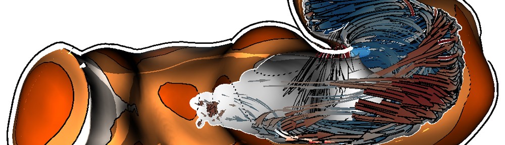
Combined 3D-display of conventional and angiographics MR data.

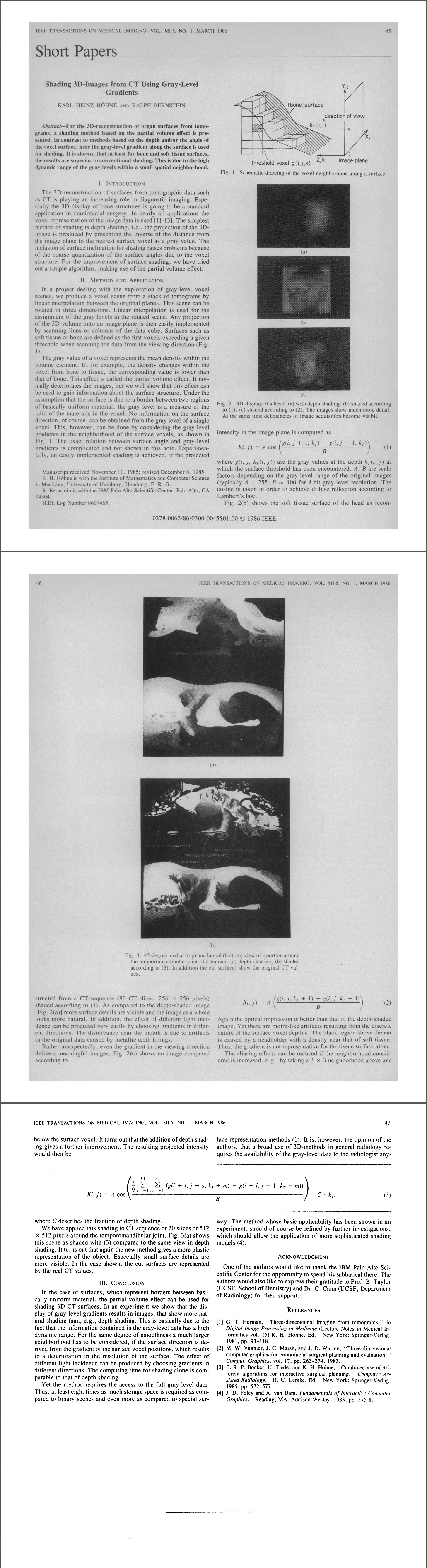
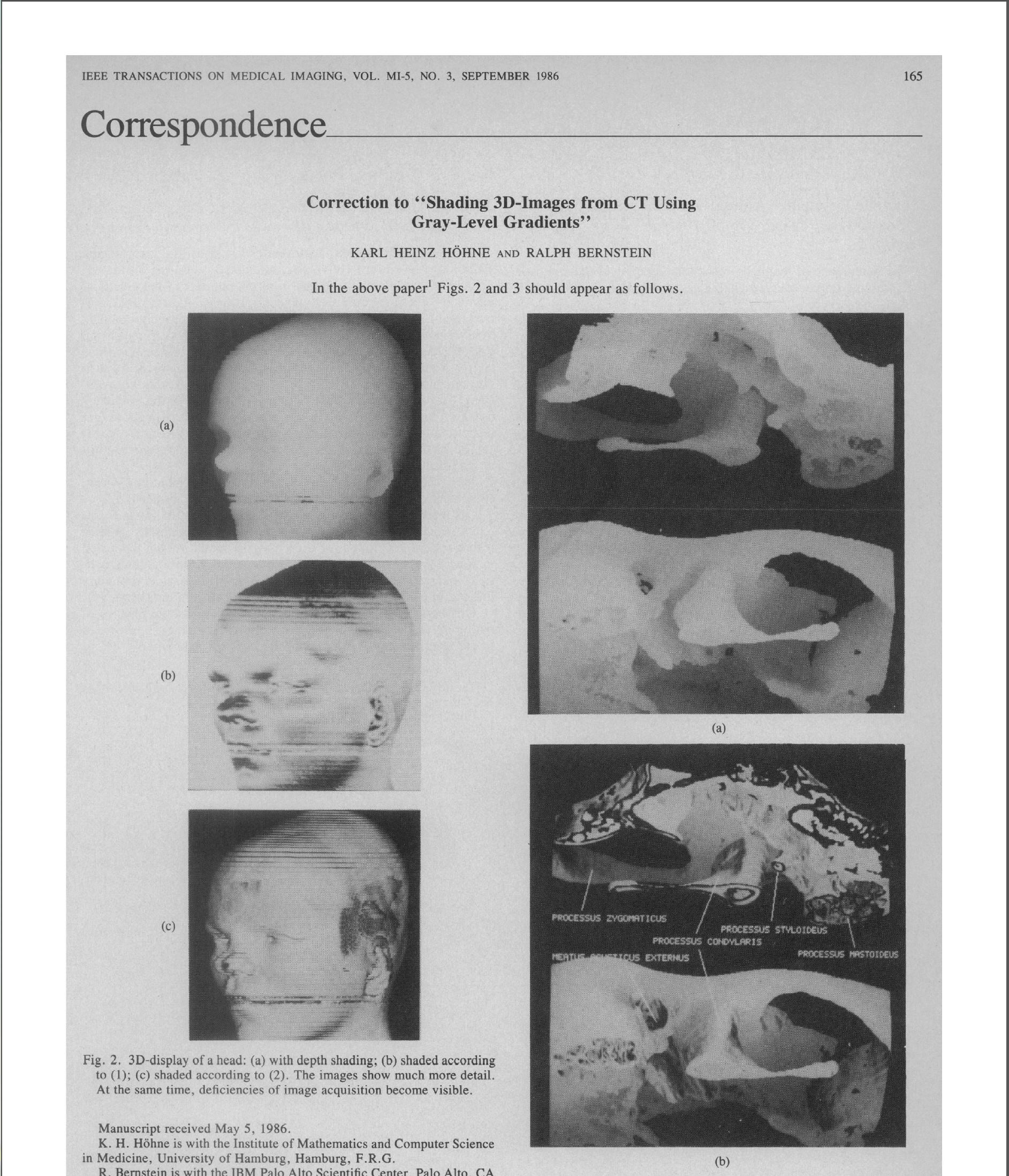
The first paper entitled ‘Shading 3D-images from CT Using Gray-Level gradients’ by Karl Heinz Höhne and Ralph Bernstein was published in 1986 in the IEEE Transactions on Medical Imaging. This paper describes a shading method based on the partial volume effect using the gray-level gradients along a surface reconstruction of CT images. What sets this method apart from current gradient vector computations is that the gray-values are sampled in screen space and not in voxel space. As a result, surfaces perpendicular to the viewer are brighter than surfaces oriented away from the viewer. In essence, this shading method simulates a headlight configuration.

The second paper entitled ‘Display of Multiple 3D-Objects Using the Generalized Voxel-Model’ by Karl Heinz Höhne, Michael Bomans, Ulf Tiede and Martin Riemer was published in 1988 in the SPIE proceedings. In this paper a generalized voxel-model for the generation of 3D-views from MR-volume data is proposed.

Using the software and hardware that was available at the time, these papers represent many contributions to the field, for instance selective volume clipping and multimodal visualization. With their work, they created a solid foundation for state-of-the-art (medical) volume rendering techniques. The two papers usually credited as first presenting the ideas behind raycasting, both published in 1988, are:

  • R. A. Drebin, L. Carpenter, and P. Hanrahan, “Volume rendering,” SIGGRAPH Comput. Graph., vol. 22, no. 4, pp. 65–74, Aug. 1988.
  • M. Levoy, “Display of surfaces from volume data,” IEEE Computer Graphics and Applications, vol. 8, no. 3, pp. 29-37, May 1988.