IEEE Visualization Contest 2010 winners announced.

This year’s IEEE Visualization Contest was medically-themed: Contestants had to demonstrate how the visualisation of multi-modal datasets could be used for neuro-surgical planning. More specifically, submissions had to show how the following two questions could be best answered:

  1. What is the relation between the lesion, functional areas and white matter tracts?
  2. How can the lesion be accessed most safely?

Recently, the winning team and three honourable mentions were announced:

Winner: Pre-Operative Planning of Brain Tumor Resections by Stefan Diepenbrock, Jörg-Stefan Praßni, Florian Lindemann, Hans-Werner Bothe and Timo Ropinski.

Honourable mention 1: An Exploration and Planning Tool for Neurosurgical Interventions by Diana Röttger, Sandy Engelhardt, Christopher Denter, Burkhard Güssefeld, Annette Hausdörfer, Gerrit Lochmann, Dominik Ospelt, Janine Paschke, QiAn Tao, Stefan Müller.

Honourable mention 2: Neurosurgical Intervention Planning with VolV by Silvia Born, Daniela Wellein, Peter Rhone, Matthias Pfeifle, Jan Friedrich, Dirk Bartz.

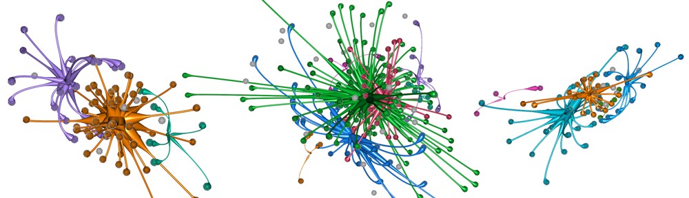
Honourable mention 3: A Fiber Navigator for Neurosurgical Planning (NeuroPlanningNavigator) by Olivier Vaillancourt, Gabriel Girard, Arnaud Bore, Maxime Descoteaux.

You can also download a 550 MB zip file with all the contributions from the contest website.

Dynamic Medical Image Visualisation at ISPA 2009

The Bergen MedViz Network is organising a special session on the Analysis and Visualization of Dynamic Images in Medical Applications at the 6th International Symposium on Image and Signal Processing and Analysis (ISPA) 2009.

Topics of interest include but are not limited to:

  • Study of perfusion using MRI, CT, US, or PET
  • Motion correction of image time series
  • Strain and stress imaging
  • Imaging of the beating heart, or the moving kidney
  • Tracking of cells, organelles, or proteins in time-lapse images
  • Compartment modeling and tracer kinetics in MRI, CT, or PET
  • Flow quantification and visualization
  • Analysis and visualization of change (e.g. displacement) in repeated image examinations
  • Image-based modeling and visualization of dynamic biological processes (e.g. diffusion)

We have good friends and colleagues in the Bergen MedViz network, so this is bound to be good.  Submit your papers!

ECR 2008

I’m currently at the European Congress of Radiology (ECR) 2008, staffing the Medical Delta booth in the Imagine section (level 2 in the Austria Centre, close to rooms X and Y), together with a number of cool people from the Erasmus Medical Centre Biomedical Imaging Group Rotterdam and the Leiden University Medical Centre Image Processing Laboratory. If you’re reading this and you’re in the neighbourhood, pop by for a chat and of course some complimentary stroopwafels and drop!

Update:

I’m back at the TU in Delft and the ECR should be finising by this afternoon. Also see the blog summaries of Marion Smits by clicking here, here and here. Below is a photo Marion took of our Medical Delta booth:

Cool Medical Delta people

Medical Visualization PhD position in Bergen, Norway

The Visualization Group at the University of Bergen in Norway currently has a job opening for a 4-year PhD research fellowship in Medical Visualization. The successful candidate will perform his/her PhD research under the supervision of Prof.dr. Helwig Hauser and Dr. Ivan Viola, two internationally respected visualization scientists.

You can apply for this exciting position via the JOBBNORGE website until March 15, 2008.

Click here for the accompanying flyer.

Illustrative Medical Visualisation

Siemens unveils MR-PET prototype

Siemens has unveiled the world’s first MR-PET scanner prototype. With this imaging system, magnetic resonance imaging (MRI) and positron emission tomography (PET) images can be acquired simultaneously. MRI supplies anatomical (structural) information as context, whilst PET yields functional data, for example on the precise location of tumours and their metastases. See the image below for an example (courtesy of Siemens):

MR-PET slices.

This is yet another new example of “native” multi-field medical datasets, and underlines the need for research on techniques that are able to visualise this type of data.

(news via MedGadget)