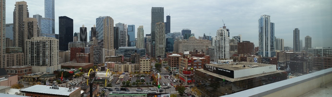
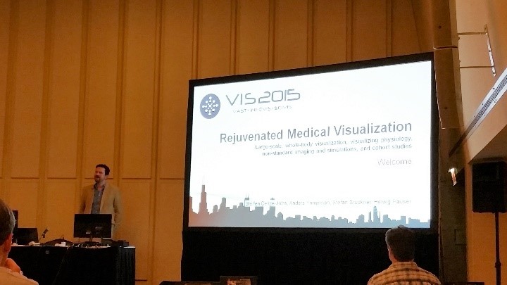
This year IEEE VIS (the conference formerly known as VisWeek) 2015 takes place in Chicago, also known as the “Windy City”. On the first day, I attended a very interesting tutorial about medical visualization entitled “Rejuvenated Medical Visualization”. This tutorial was opened by Steffen Oeltze-Jafra, the organizer of this event. He welcomed the audience and introduced the other speakers in the tutorial: Anders Ynnerman, Stefan Bruckner and Helwig Hauser.
I was very happy to see that in 2013, the IEEE Vis conference hosted again a separate session on biomedical visualization. On top of the five talks given in this session, five more interesting talks, also related to MedVis, were distributed over the conference program. Before the event started, I considered it a good omen that the inwards of the conference hotel looked like a gigantic corpus with the conference attendees accommodated along the costal arches.
The session on biomedical visualization was opened by Jan Kretschmer from the FAU Erlangen and Siemens Healthcare Computed Tomography, Forchheim, Germany. He gave a talk on the interactive patient-specific modeling of vasculature by means of sweep surfaces. He showed how vascular segmentations may be polished in a fast, interactive, and intuitive way such that high-precision models for blood flow simulations are generated on the fly. The modeling approach is robust, eligible for clinical on-site application, and it delivers smooth high quality results.
Xin Zhao from Stony Brook University presented a novel area-preservation mapping/flattening method using the optimal mass transport technique. Compared to previous methods, the size and area of each fold component are preserved facilitating quantitative analyses. Two interesting and very relevant applications from a medical point of view were presented: brain surface flattening and colon flattening. In the former, the correct detection and quantification of brain folds is crucial. Traditional approaches induce severe area distortions and therefore hamper these tasks. In colon flattening, the detection and measurement of polyps benefit from the new method.
A tailor-made algorithm for colon flattening was presented by Krishna Chaitanya Gurijala from Stony Brook University. In contrast to previous approaches, the algorithm is shape-preserving and robust to topological noise. It dispenses denoising the data as a pre-processing step and instead replaces the original Euclidean metric of the colon surface with a heat diffusion metric that is insensitive to topological noise. Virtual colonoscopy greatly benefits from the new approach since shape and area of polyps are preserved.
Johanna Beyer from Harvard University, Cambridge (previously with the King Abdullah University of Science and Technology (KAUST), Saudi Arabia) concluded the biomedical session. She presented a system for the query-guided visual analysis of large volumetric neuroscience data: the ConnectomeExplorer. The system facilitates the integrated visual analysis of volume data, segmented objects, connectivity information, and additional meta data. Powerful query algebra allows neuroscientists to pose domain-specific questions on the data in an intuitive manner. Johanna’s presentation was completed by an impressive demonstration of the systems performance in typical use-case scenarios.
Moritz Ehlke from the Zuse Institute Berlin presented an approach to render virtual X-ray projections of deformable tetrahedral meshes that runs very fast on the GPU. The purpose of generating these projections is the reconstruction of 3D anatomy from a single or a few 2D X-ray images. In an iterative optimization process, the tetrahedral mesh of a statistical shape and intensity model of an anatomical structure is transformed, such that it represents plausible candidates for a patient-specific shape and density distribution. Each transformation result is then converted to a virtual X-ray projection, whose X-ray attenuation is finally compared to the clinical 2D X-ray. The best candidate provides a plausible representation of 3D anatomy which was demonstrated for the pelvic bone.
Bret Jackson from the University of Minnesota presented a prop-based, tangible interface for 3D interactive visualization of thin fiber structures. He demonstrated the exploration of fiber orientations in second-harmonic generation microscopy of collagen fibers by means of a paper prop, a depth sensing camera, and a low-cost 3D display. The paper prop is tracked and the visualization is restricted to fibers oriented in the direction specified by the prop, i.e. the user. Different gestures, one- and two- handed, are supported for filtering fibers, adjusting the fiber similarity threshold, slicing the volume, and rotating or rolling the volume.
Benjamin Köhler from the Otto-von-Guericke University Magdeburg, Germany gave a talk on the semi-automatic vortex extraction in 4D PC-MRI cardiac blood flow data by means of line predicates. The relation of blood flow patterns, e.g., vortices, and vascular pathologies is currently a hot topic in cardiovascular research. Benjamin compared various vortex extraction methods to determine the most suitable one for cardiac blood flow. He integrated several dedicated flow visualization techniques and the vortex computation in a system that is fully implemented on the GPU to provide real-time feedback. The system was demonstrated based on ten datasets with different pathologies like coarctations, Tetralogy of Fallot and aneurysms and evaluated at the Heart Center Leipzig. A video is available here.
Adrian Maries from the University of Pittsburgh presented GRACE: A visual comparison framework for integrated spatial and non-spatial geriatric data. These high-dimensional data span volumetric images and variables such as age, gender or walking speed. Their concurrent analysis is supported by a multiple coordinated view system comprising volume rendering panels, dendogram panels, and a Kiviat graph. Techniques from statistics are integrated to quantify potential neurology-mobility connections. The usefulness of the framework for generating and refining hypotheses was demonstrated on two case studies. In the paper, the authors report their lessons learned from designing visualizations for concurrently analyzing spatial and non-spatial data. Check the videos here.
Thomas Schultz from the University of Bonn, Germany gave a very good talk on the application of spectral clustering to medical image analysis. He showed a system that makes this powerful and versatile technique more accessible to users via an open-box approach, in which an interactive system visualizes the involved mathematical quantities, suggests clustering parameter values, and provides immediate feedback to support the required decisions, e.g., on the number of clusters. The system further supports the filtering of outliers and the recording of user actions and their translation to other data containing the same structures. Thomas demonstrated the system based on chest CT and brain MRI data.
This year, IEEE VisWeek hosted no special session dedicated to medical visualization but
five contributions to this field were spread over the conference program.
Jian Chen from the University of Maryland gave a talk on how stereo and screen size effect the legibility of three-dimensional streamtube visualizations. The effects were studied in the context of visually exploring dense fiber tracts reconstructed from diffusion magnetic resonance imaging data. A user study comprising 12 participants who had to perform five different tasks, e.g., find the endpoints of fiber tracts and judge if tracts belong to the same bundle, was carried out. In contrast to the initial hypotheses of Chen and
colleagues, completion time did not improve by using a larger display and performance accuracy was even hurt by introducing stereo. See the paper for further exploration of the results [1].
Rocco Gasteiger from the University of Magdeburg, Germany gave a compelling talk on the automatic detection and visualization of qualitative hemodynamic characteristics in cerebral aneurysms. He focused on the so-called inflow jet and the impingement zone, both being characteristics which are correlated with the risk of aneurysm rapture. Special care was taken in generating expressive visualizations of the detected features by means of glyphs, texture, Fresnel shading, and surface contours. The work was rounded off by a user study involving six domain experts who show high interpersonal variance in manually specifying the features but agreed on the good value of the presented visualizations [2]. A supplemental video can be watched here:
Markus Hadwiger from the King Abdullah University of Science and Technology, Saudi Arabia presented the first volume visualization system that scales to petascale volumes imaged as a continuous stream of high-resolution electron microscopy images. Markus and his team developed the system in collaboration with neuroscientists who wish to analyze brain function by measuring large blocks of brain tissue. The system can accept a constant stream of 2D image tiles from a microscope thereby handling missing data naturally and avoiding the expensive computation of any 3D multi-resolution representation such as an octree. A novelty of the system is that most computations are restricted to the currently visible volume data [3]. Watch the accompanying video here:
Alexander Bock from the Linköping University, Sweden presented the ray-casting of high-order finite element (FE) models for visualizing and analyzing strain of the human heart muscle. A straightforward ray-casting approach is inadequate for interactive data exploration due to the computational complexity of transforming the sample points along each ray into the non-uniform grid of the FE model. Hence, Alexander and his colleagues decoupled the expensive transformation from the rendering stage by means of proxy rays cast in FE space, thereby allowing it to be performed within a precomputation stage. The nice work was rounded off by an analysis of the error that is introduced by the presented approach [4].
Gunnar Läthén, also from the Linköping University, Sweden, gave a talk on improving transfer functions for volume rendering blood vessels in computed tomography angiography (CTA) data [5]. These data are acquired by means of injecting a contrast agent, which leads to an enhancement of the vessels. Due to variations in mixture concentration of contrast agent in the blood stream, the enhancement varies locally and transfer function presets often do not yield optimal images. Hence, Gunnar and his colleagues propose an automatic, optimization-based method that shifts transfer function presets to account for general deviations and local variations of the intensity of contrast enhanced blood vessels. The method is illustrated for clinically relevant CT angiography datasets.
(We are happy and grateful that Dr. Steffen Oeltze from the University of Magdeburg Visualization Group could write this short report on the medical visualization session and other medvis-related papers at IEEE VisWeek 2011.)
This year, the IEEE VisWeek has been completed by an excellent session on medical visualization hosting five contributions from three European countries. Roy van Pelt gave a compelling talk on the exploration of cardiovascular 4D MRI blood-flow using stylistic visualizations. His comic-inspired illustrative glyphs coupled with timelines outperform traditional particle renderings. Interactive virtual probing of the flow
avoids a tedious segmentation process in qualitative inspection.
Rostislav Khlebnikov presented a new approach to tumor accessibility planning. It exploits a well-known natural phenomenon related to light scattering at dust particles which is also called crepuscular rays. In the generated 2D/3D images, light beams in different colors that shine through the skin indicate the access paths and their associated risk.
Christian Dick presented new visualization techniques for conveying distances in interactive 3D implant planning. The design of very intuitive distance glyphs and colored slice sets was completed by a carefully accomplished, convincing user study.
Rocco Gasteiger introduced the FlowLens for focus+context visualization of blood flow in cerebral aneurysms. It supports an exploration of certain hemodynamic attributes in the lens region within the context of other attributes thereby avoiding the cognitive effort involved in mental superimposition of side-by-side visualizations. Please watch the supplemental video:
The session was completed by the interesting talk of Artem Amirkhanov on the reduction of metal artifacts in industrial 3D X-ray CT images. He presented a projection-space pipeline in which metal is separated from the other materials before projection and then fused again with the initial reconstruction after projection.
Other talks not being part of the session but also related to medical visualization were given by Claes Lundström on the application of a multi-touch table system to orthopedic surgery planning, Christian Rieder on real-time approximation of the ablation zone for radiofrequency ablation (see the very nice video), Joseph Marino on context preserving maps of tubular structures, e.g., the colon, and Paolo Angelelli on straightening aortic blood flow for side-by-side visualization.