Visualising Medical Heritage and Virtual Reality
Thursday 23rd May, 6.30pm
Our Visualising Medical Heritage Project brings medical history to life and enables enhanced access to our museum collection through cutting edge visualisation technology. Join us for a series of events where we will explore the different potentials of visualisation, combining medical history, science and the creative arts.
This workshop looks at the wonderful world of VR. From 3D digital modelling to VR headsets and Minecraft, find out how new technologies are helping us to bring our medical heritage to life.
We’ll also be joined by Holoxica Ltd and The School of Simulation and Visualisation at the Glasgow School of Art – they’ll be demonstrating some amazing, new technologies which explore imaginative uses of 3D digital visualisation and interaction technologies.
The event starts at 6:30pm and is suitable for adults and children 12+
Venue: Royal College of Physicians and Surgeons of Glasgow
Book here – https://rcpsg.ac.uk/events/HERITAGEVMH230519-2019-05-23-121
Our Visualising Medical Heritage project is supported by Museums Galleries Scotland.
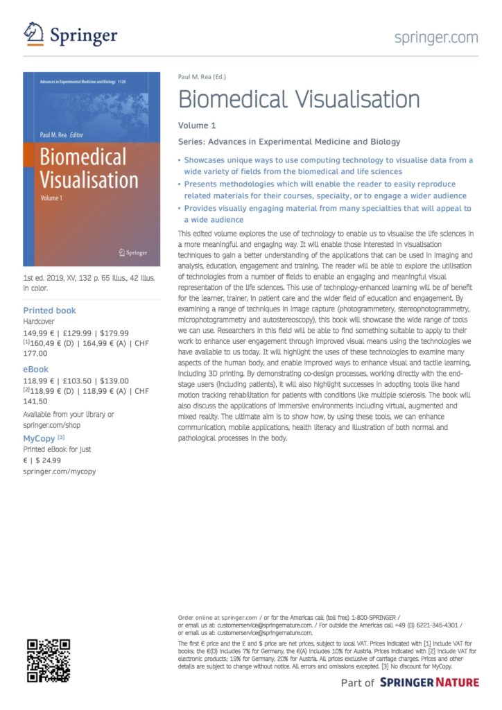
The first ever volume of Biomedical Visualisation is out now. We have several more volumes to come out over the coming months, so keep an eye out for that.
I have an exciting opportunity to contribute a book chapter to the book volume entitled Biomedical Visualisation, Springer International Publishing, of which I am Editor. This will be within the Advances in Experimental Medicine and Biology series. I am looking for those involved in using technologies to improve our understanding of the human body, related biological processes and educating via technological methodologies. Volume 1 and 2 are out soon. Contact me if interested with a proposal.
The Eurographics Association organizes a biannual competition to acknowledge the contribution of computer graphics in the medical field, and to encourage further development. Originally called “Eurographics Medical Prize”, the competition was renamed to “Dirk Bartz Prize for Visual Computing in Medicine” in 2010 — in honor of Dirk Bartz who passed away far too early in March 2010. Dirk Bartz was a highly
recognized and enthusiastic scientist, teacher and promoter of Visual Computing in Medicine.
Submissions to the Dirk Bartz Prize for Visual Computing in Medicine 2019 are being invited from researchers and developers who can demonstrate that a particular benefit in a medical application has resulted from the use of visual computing technology that they have produced/developed. Submissions from all areas of visual computing — examples include the use of new data visualization techniques, interaction methods, or virtual/augmented environments are welcome. Entries typically summarize a body of research and/or development that has been conducted over the course of a project, PhD thesis, etc. and particular weight is put on demonstrating the medical impact of the work.
The submission deadline for next year’s award is January 25, 2019. Please find more information on the official webpage.
We have a very exciting public engagement (adult only) event called Night at the Museum of Anatomy – Past, Present and Future. It will be held from 18:00-20:00 on the 27th September at the University of Glasgow with partners from the Royal College of Physicians and Surgeons of Glasgow, the School of Simulation and Visualisation, The Glasgow School of Art, the NHS and featuring anatomical, historical and pathology specimens from our Anatomy Facility, as well as body painting, at the University of Glasgow. Click HERE to find out more and be sure to register fast to guarantee your place. It is set to be a very popular event!
Watch our Invited Speaker David Sime, talk about digital technologies from a marketing perspective in education. This was part of the 11th Annual Learning and Teaching Conference at the University of Glasgow, 28th and 29th March 2018.
Watch the full Keynote by Professor Paul Chapman, Head of the School of Simulation and Visualisation, The Glasgow School of Art, talking about “The Pros & Cons of VR for Learning & Teaching”. This was part of the 11th Annual Learning and Teaching Conference at the University of Glasgow, 28th and 29th March 2018.
Update: The submission deadline has been extended to March 4th due to popular demand!
The “call for papers” for the EuroRVVV (EuroVis Workshop on Reproducibility, Verification, and Validation in Visualization) was released yesterday and this year it is focused on medical visualization! The topic is “From Medical Visualization Concepts to Certified Applications”. We hope that everyone gets excited about it and submits as many papers as possible until February 26th, 2016!
I was very happy to see that in 2013, the IEEE Vis conference hosted again a separate session on biomedical visualization. On top of the five talks given in this session, five more interesting talks, also related to MedVis, were distributed over the conference program. Before the event started, I considered it a good omen that the inwards of the conference hotel looked like a gigantic corpus with the conference attendees accommodated along the costal arches.
The session on biomedical visualization was opened by Jan Kretschmer from the FAU Erlangen and Siemens Healthcare Computed Tomography, Forchheim, Germany. He gave a talk on the interactive patient-specific modeling of vasculature by means of sweep surfaces. He showed how vascular segmentations may be polished in a fast, interactive, and intuitive way such that high-precision models for blood flow simulations are generated on the fly. The modeling approach is robust, eligible for clinical on-site application, and it delivers smooth high quality results.
Xin Zhao from Stony Brook University presented a novel area-preservation mapping/flattening method using the optimal mass transport technique. Compared to previous methods, the size and area of each fold component are preserved facilitating quantitative analyses. Two interesting and very relevant applications from a medical point of view were presented: brain surface flattening and colon flattening. In the former, the correct detection and quantification of brain folds is crucial. Traditional approaches induce severe area distortions and therefore hamper these tasks. In colon flattening, the detection and measurement of polyps benefit from the new method.
A tailor-made algorithm for colon flattening was presented by Krishna Chaitanya Gurijala from Stony Brook University. In contrast to previous approaches, the algorithm is shape-preserving and robust to topological noise. It dispenses denoising the data as a pre-processing step and instead replaces the original Euclidean metric of the colon surface with a heat diffusion metric that is insensitive to topological noise. Virtual colonoscopy greatly benefits from the new approach since shape and area of polyps are preserved.
Johanna Beyer from Harvard University, Cambridge (previously with the King Abdullah University of Science and Technology (KAUST), Saudi Arabia) concluded the biomedical session. She presented a system for the query-guided visual analysis of large volumetric neuroscience data: the ConnectomeExplorer. The system facilitates the integrated visual analysis of volume data, segmented objects, connectivity information, and additional meta data. Powerful query algebra allows neuroscientists to pose domain-specific questions on the data in an intuitive manner. Johanna’s presentation was completed by an impressive demonstration of the systems performance in typical use-case scenarios.
Moritz Ehlke from the Zuse Institute Berlin presented an approach to render virtual X-ray projections of deformable tetrahedral meshes that runs very fast on the GPU. The purpose of generating these projections is the reconstruction of 3D anatomy from a single or a few 2D X-ray images. In an iterative optimization process, the tetrahedral mesh of a statistical shape and intensity model of an anatomical structure is transformed, such that it represents plausible candidates for a patient-specific shape and density distribution. Each transformation result is then converted to a virtual X-ray projection, whose X-ray attenuation is finally compared to the clinical 2D X-ray. The best candidate provides a plausible representation of 3D anatomy which was demonstrated for the pelvic bone.
Bret Jackson from the University of Minnesota presented a prop-based, tangible interface for 3D interactive visualization of thin fiber structures. He demonstrated the exploration of fiber orientations in second-harmonic generation microscopy of collagen fibers by means of a paper prop, a depth sensing camera, and a low-cost 3D display. The paper prop is tracked and the visualization is restricted to fibers oriented in the direction specified by the prop, i.e. the user. Different gestures, one- and two- handed, are supported for filtering fibers, adjusting the fiber similarity threshold, slicing the volume, and rotating or rolling the volume.
Benjamin Köhler from the Otto-von-Guericke University Magdeburg, Germany gave a talk on the semi-automatic vortex extraction in 4D PC-MRI cardiac blood flow data by means of line predicates. The relation of blood flow patterns, e.g., vortices, and vascular pathologies is currently a hot topic in cardiovascular research. Benjamin compared various vortex extraction methods to determine the most suitable one for cardiac blood flow. He integrated several dedicated flow visualization techniques and the vortex computation in a system that is fully implemented on the GPU to provide real-time feedback. The system was demonstrated based on ten datasets with different pathologies like coarctations, Tetralogy of Fallot and aneurysms and evaluated at the Heart Center Leipzig. A video is available here.
Adrian Maries from the University of Pittsburgh presented GRACE: A visual comparison framework for integrated spatial and non-spatial geriatric data. These high-dimensional data span volumetric images and variables such as age, gender or walking speed. Their concurrent analysis is supported by a multiple coordinated view system comprising volume rendering panels, dendogram panels, and a Kiviat graph. Techniques from statistics are integrated to quantify potential neurology-mobility connections. The usefulness of the framework for generating and refining hypotheses was demonstrated on two case studies. In the paper, the authors report their lessons learned from designing visualizations for concurrently analyzing spatial and non-spatial data. Check the videos here.
Thomas Schultz from the University of Bonn, Germany gave a very good talk on the application of spectral clustering to medical image analysis. He showed a system that makes this powerful and versatile technique more accessible to users via an open-box approach, in which an interactive system visualizes the involved mathematical quantities, suggests clustering parameter values, and provides immediate feedback to support the required decisions, e.g., on the number of clusters. The system further supports the filtering of outliers and the recording of user actions and their translation to other data containing the same structures. Thomas demonstrated the system based on chest CT and brain MRI data.
Yesterday, Christian Rieder of Fraunhofer MEVIS successfully defended his Ph.D. thesis entitled Interactive Visualization for Assistance of Needle-Based Interventions at the Jacobs University Bremen.Supervised by Horst Hahn, Christian made a number of significant contributions in the last years, leading to strong publications at VisWeek and EuroVis, and a MedVis-Award distinction in 2010. Thus, it was not too surprising that the thesis was assessed with the highest possible grade.
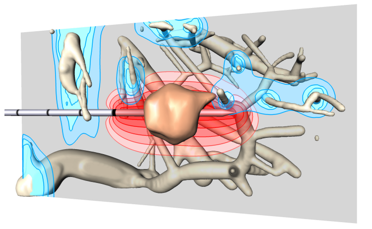
Visualization from Christian’s 2011 VisWeek paper showing RF applicator, tumor, the approximated ablation zone in red and thermal cooling of blood vessels in blue.
Christian’s work aims at supporting clinical workflows, primarily in radio frequency ablation, supporting both the pre-interventional as well as the interventional stage with highly advanced and carefully adapted visualizations indicating tumors, risk structures, security margins as well as results from approximative simulations that predict the thermal lesion produced by RFA. Illustrative techniques, smart map projections, very efficient GPU realizations as well as careful evaluations with relevant physicians are landmarks of Christian’s work, which may be explored on his website in detail.
(editor: Thanks to Prof. Bernhard Preim for submitting this news. We have always been a fan of Christian and his work, and we are very happy to hear of this success!)