Call for Participation: MedVis-Award 2016

The 7th “Karl-Heinz-Höhne Award for Visualization in Medicine” (in short medvis-award) is now accepting submissions. Besides eternal glory, the top contestants of this biannual competition will receive in total 1.000 EUR, donated by BrainLAB AG. You can only apply if you are a young scientist with a diploma thesis or with up to two publications (published or to be reviewed) in the field of medical visualization. Does this sound like you or someone you know? Find out more about the award here and check out previous winners here.

The submission deadline is the 4th of May (May the 4th be with you!) and the lucky winners will be receiving their award at VCBM 2016 in Bergen, Norway 🙂

Announcing VCBM & MedViz 2016!

It’s no secret the Eurographics Workshop on Visual Computing for Biology and Medicine (VCBM) is one of my favorite events for some years already, all medical visualization all the time! Since last year, it turned into an annual workshop, which means we get to enjoy another VCBM in 2016 already, from the 7th of September until the 9th. This edition promises to be extra epic for several reasons, outlined below:

  1. It will be in Bergen, Norway. Bergen, for those of you that never visited, is a truly amazing city situated between majestic mountains and a beautiful harbor. You could do worse! “The Gateway to the Fjords of Norway”, people!
  2. Additionally, also speaking from personal experience, the Bergen Vis group is filled with awesome people that do great research.
  3. If this is still not convincing you of the epicness, next year the 10th annual Medviz conference will be held at the same time!

‘So where do I sign up?’ I hear you thinking. The official website describing all the details is here, there is a Facebook page for you to like (if not love!) here and even a Facebook event here!

Conference Report: EG VCBM 2015 Chester (UK)

Recently I had the pleasure of attending the Eurographics Workshop on Visual Computing for Biology and Medicine (VCBM) 2015 conference for the third and potentially, but not hopefully, last time. This year it was held in Chester (UK) at The Riverside Innovation Centre at the University of, you guessed it, Chester! In this conference report I will summarize some personal highlights. Repeating last year’s tradition, I again tweeted a picture for almost every talk. I still don’t think Twitter is really gaining traction among the scivis community, and I wonder what it would take to change it (or if it even needs to change ^^). As every year, given the theme of the conference almost every talk is relevant to our medical visualization interests, but I would like to briefly summarize only a couple of them here. Check the full list of papers and posters presented here if this is not enough to satiate your VCBM-craving-needs. Continue reading

VCBM 2015 Chester (UK) call for papers and posters: Deadlines imminent!

The “Call for Papers and Posters” for VCBM (AKA Eurographics Workshop on Visual Computing for Biology and Medicine) 2015 was released a while ago. As you probably already know, VCBM is an excellent venue for medical visualization work and this year it will be held in Chester, UK.

The deadline for full paper submission is June 21st and the posters need to be submitted by August 7th. Don’t miss this opportunity to present your work at this excellent location and please take a look at the website for more details.

Conference Report: EG VCBM 2014 Vienna (Austria)

In the spirit of better late than never, the Eurographics Workshop on Visual Computing for Biology and Medicine (VCBM) 2014 conference report, summarizing some personal highlights. At VCBM 2014, we tweeted a picture for every talk and then some, which was arguably a bit much, but still a lot of fun. The VCBM organization also posted a great recap using Storify at their website. Given the theme of the conference, almost every talk is relevant to our medical visualization interests, but I would like to briefly summarize only a couple of them here. The benefit of delaying so long in writing this is that there are a lot of videos online by now. I will try to let the videos speak a 1000 words where available instead of getting too verbose. Onwards to the highlights!

The venue was really amazing, VCBM was held in the Universitätscampus
Altes AKH, but not just in any old lecture hall, it was the former anatomical theatre of the AKH and still has the original marble slab that was used as a dissection table:

Hörsaal D at the AKH

Hörsaal D at the AKH

A day before the start of VCBM itself, the VCBM fachgruppe (working group) had a meeting with six interesting talks. This was followed by a social event, a guided tour of the Narrenturm. Built in 1874 to treat mental patients, it now serves as a museum for the Pathologic-Anatomical Collection. The Narrenturm features a huge collection of moulages. These are wax models of diseases made based on real patients and used in medical education, which is cool and slightly creepy at the same time. This tour was followed up by a delicious dinner at Unibräu for those who didn’t lose their appetite after what they had seen during the tour.

On Thursday, VCBM itself kicked off with an opening by Katja Bühler. After this we enjoyed a keynote by Anna Vilanova on the future of medical visualization. Anna presented medical visualization as a field that is between fields: computer graphics and medical imaging. She talked us through the past, present and future of medvis and  going from facilitating analysis of the known to unraveling the unknown using visualization. A memorable quote from her talk:

“If the brain were so simple that we could understand it, we would be so simple we couldn’t” – Lyall Watson

Thursday featured four interesting sessions on Multivariate Data Analysis, Segmentation and Uncertainty, Microscopy and Visual Analytics for Biology:

  • Multivariate Data Analysis:
  • Segmentation and Uncertainty:
    • Peter Faltin presented his work on “Extracting and Visualizing Uncertainties in
      Segmentations from 3D Medical Data” [3]. He introduces a new processing chain comprising a series of carefully selected and well-matched steps to
      determine and visualize a segmentation boundary. Additionally, a novel visualization method was presented, specifically designed to simultaneously provide information about 3D morphology, confidence and possible errors.
  • Microscopy:
  • Visual Analytics for Biology:
    • I really enjoyed the talk by Nicolas Swoboda on “Visual and Quantitative Analysis of Higher Order Arborization Overlaps for Neural Circuit Research” [5]. The overlaps they are reffering to, consist of two or more neurons and indicate a potential anatomical connection. They present a novel tool for potential connectivity exploration by providing for the first time the possibility to compute and visualize higher order arborization overlaps on the fly (for fruit fly brains, well played!) and to interactively explore this information in its spatial
      anatomical context and on a quantitative level. Slides of the talk are available here and this is the accompanying video:

In the evening we hiked up through the vineyards of Vienna to the main social event: dinner at the Waldgrill Cobenzl. The view on the vineyards and Vienna itself was really stunning. We enjoyed a delicious buffet dinner accompanied by Sturm. Sturm is grape juice that has just started fermenting and is only available for a limited time every year, so we were lucky VCBM was held in Vienna exactly during Sturm time. After dinner the winners of the Karl-Heinz Höhne Award for Medical Visualization were announced:

I would love to tell you who the winner’s were, but the official announcement has not been made yet ;), so I don’t dare… Congratulations to the award winnners nonetheless, you know who you are ^^.

On the second and last day of VCBM there were sessions on Volume Visualization, Image Registration and Data Reconstruction for Medical Interventions,  Visual Explanations and Display Techniques as a keynote by Nigel John entitled ‘Visual Computing in Healthcare – from the Research Lab into the Hospital”. In the keynote he presented several case studies and discussed some of the challenges
involved in deploying visual computing solutions in a hospital setting.

VCBM wrapped up with the awards ceremony. The best paper award went to:

The honorable mentions can be found here. Our congratlations to the authors! Ivan Viola closed the conference and announced the location for next year (this year by now ^^): VCBM 2015 will be held in Bangor (UK) and will from now on be an annual workshop instead of bi-annual (once every two years, not the twice every year-type). To conclude this summary, I’d really like to thank the organizers of this excellent workshop. Interesting talks, a beautiful location, good food, great people once again!

References:

  • [1]: Robust Cardiac Function Assessment in 4D PC-MRI Data. Köhler, Benjamin; Preim, Uta; Gutberlet, Matthias; Fischbach, Katharina; Preim, Bernhard
  • [2]: The iCoCooN: Integration of Cobweb Charts with Parallel Coordinates for Visual Analysis of DCE-MRI Modeling Variations. Raidou, Renata; Breeuwer, Marcel; Vilanova, Anna
  • [3]: Extracting and Visualizing Uncertainties in Segmentations from 3D Medical Data. Faltin, Peter; Chaisaowong, Kraisorn; Kraus, Thomas; Merhof, Dorit
  • [4]: Interactive Labeling of Toponome Data. Oeltze-Jafra, Steffen; Pieper, Franz; Hillert, Reyk; Preim, Bernhard; Schubert, Walter
  • [5]: Visual and Quantitative Analysis of Higher Order Arborization Overlaps for Neural Circuit Research. Swoboda, Nicolas; Moosburner, Judith; Bruckner, Stefan; Yu, Jai Y.; Dickson, Barry J.; Bühler, Katja
  • [6]: Visibility-Driven Processing of Streaming Volume Data. Solteszova, Veronika; Birkeland, Åsmund; Viola, Ivan; Bruckner, Stefan
  • [7]: Towards Clinical Deployment of Automated Anatomical Regions-Of-Interest. Lindholm, Stefan; Forsberg, Daniel; Ynnerman, Anders; Knutsson, Hans; Andersson, Mats; Lundström, Claes
  • [8]: RegistrationShop: An Interactive 3D Medical Volume Registration System. Smit, Noeska; Klein Haneveld, Berend; Staring, Marius; Eisemann, Elmar; Botha, Charl; Vilanova, Anna
  • [9]: Survey of Labeling Techniques in Medical Visualizations. Oeltze-Jafra, Steffen; Preim, Bernhard

VCBM 2014 Vienna (Austria) – Call for papers and posters!

The “Call for Papers and Posters” for VCBM (AKA Eurographics Workshop on Visual Computing for Biology and Medicine) 2014 has been released! VCBM is an excellent venue for medical visualization work and this year it will be held in Vienna, Austria.

The deadline for full paper submission is June 19 and the posters need to be submitted by August 7. Don’t miss this opportunity to present your work at this excellent location and please take a look at this website for more details. In other news: did you know our website also features a conference calendar that you can add to your personal calendar?

vcbm2014

Start submitting those papers and posters for VCBM 2014!

Call for Participation: MedVis-Award 2014

The 6th “Karl-Heinz-Höhne Award for Visualization in Medicine” (in short medvis-award) is now accepting submissions. You can only apply if you are a young scientist (m / f) with a diploma thesis or with up to two publications (published or to be reviewed) in the field of medical visualization. Does this sound like you or someone you know? Find out more about the award here and check out last year’s winners here.

The submission deadline is the 4th of May and the lucky winners will be receiving their award at VCBM 2014 in Vienna, Austria.

vcbm2014

The award ceremony will be held at VCBM 2014

medvis.org 2012 summary

First of all, happy new year from all of us at medvis.org! May 2013 bring you many job opportunities, research successes and/or good medical visualizations. I’d like to start the new year by looking back at last year briefly. I’ve taken a look at some of our blog statistics and will provide a short summary for those interested below:

EG VCBM 2012 Norrköping (Sweden) Report

I started writing this post in the train from Norrköpping back to Stockholm Arlanda airport after visiting the truly excellent EG VCBM 2012 (Eurographics Workshop on Visual Computing for Biology and Medicine). While enjoying the beautiful view on the stunning Swedish landscape that features many lakes, trees with autumn leaves and a nice autumn sun, I briefly summarized the conference highlights. Please note that these were the conference highlights for me personally, and are not necessarily a reflection of the actual conference highlights. There were so many great talks, I lost count, but in the interest of not making this blog post drag on for too long, I’ll restrict myself to just briefly summarizing a couple of them here.

The first day started with a word of welcome from the chairs Timo Ropinski and Anders Ynnerman and an excellent keynote by Anders Persson (Director of the CMIV): ‘Visualization of Quantified Medical Image Data – Key to the Future?’. While showing us beautiful datasets acquired from Dual Energy CT (DECT), he stressed the importance of making quantified imaging data usable in clinical practice. This can be achieved by working in close collaboration with medical centers to make sure the techniques we are developing are usable by and useful to clinicians or medical researchers.

Dual-energy CT (DECT) with two X-ray sources running simultaneously at different energies allows obtaining additional information about the elementary chemical composition of computer tomography scanned material [1].

The first session of the first day, Tractography and Connectivity, featured a talk by Anne Berrescalled ‘Tractography in Context: Multimodal Visualization of Probabilistic Tractograms in Anatomical Context’ [2]. Her approach is a way of handling the visibility issues that arise when presenting probabilistic tractograms within anatomical context. Probabilistic tractography data is presented in a ‘glass brain’ rendering that provides anatomical context together with an MRI slice plane, in a collaboration with neurological domain experts.

Tractography data in anatomical context [2].

In the Ultrasound session, the talk by Daniel Tenbrinck entitled ‘Impact of Physical Noise Modeling on Image Segmentation in Echocardiography’ really stood out for me [3]. Not only was it presented extremely skillfully, but the authors made a convincing argument against the frequently used Gaussian noise model assumption and demonstrated the positive effect of chosing a more suitable model such as the Loupas model. In this same session, Veronika Solteszova and 
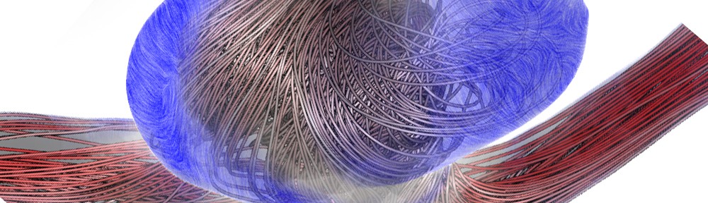
Linn Emilie Saevil Helljesenpresented a new way of filtering 3D ultrasound using lowest-variance streamlines that reduces noise in 3D ultrasound datasets with impressive results [4].

Raw 3D ultrasound scan on the left and a visualization of the same dataset filtered with the lowest-variance streamline method [4].

The final session of the day, on Multimodality included a great talk by Florian Weilerpresenting his work ‘On the Value of Multi-Volume Visualization for Preoperative Planning of Cerebral AVM Surgery’ [5]. In the surgical treatment of cerebral arteriovenous malformations (AVMs), thorough preoperative planning is required using information about the arteries and veins of the lesion from different image sets. The authors merge these image sets and visualize them in an interactive application.

Multi-Volume Visualization for Preoperative
Planning of Cerebral AVM Surgery [5].

Before making our way to the conference dinner, we were given a very special tour of the universe by Anders Ynnerman. I’m typically not at all interested in space stuff, but this was something else! Anders actually showed us the entire universe in the Dome, using real data from NASA. Besides the visually stunning features of this tour, Anders managed to make it a truly amazing experience by providing commentary full of interesting anecdotes and impressive facts. The feeling that you get when experiencing all this is very hard to describe, so I will not even try. But I will say this though, if you’re ever in the area and get the chance to see this, go for it!

On the second conference day, the Segmentation and Simulation session’s second talk presented by Frank Heckel ‘Sketch-based Image-Independent Editing of 3D Tumor Segmentations Using Variational Interpolation’ was also very interesting [6]. By allowing the user to intuitively draw adjustments on single slices, automatic segmentations can easily be adjusted by medical experts. This is currently shown on CT, but since the adjustments are not image-based, the technique is valid for arbitrary modalities.

Sketch-based segmentation editing: (a) initial segmentation (yellow), manual correction (blue) and (b) 3D result after editing with our variational-interpolation-based approach [6].

After this session, the Visual Computing Systems session started with the impressive ‘Visually Guided Mesh Smoothing for Medical Applications’ talk by Tobias Moench [7]. The authors provided a way of interactively smoothing a mesh by trying out several parameters and simultaneously show the effect of these settings on mesh quality by using a GPU mesh smoothing implementation. By calculating the model quality for several parameter combinations, an optimal set of smoothing parameters can be automatically suggested as well (click to see a cool realtime mesh smoothing video).

The medvis.org overlord who also happens to be my supervisor, Charl Botha, presented ‘BrainCove: A Tool for Voxel-Wise fMRI Brain Connectivity Visualization’ in the same session [8]. I am obviously biased here, but I thought it was an interesting and entertaining presentation. I mean it featured the BrainCleaver, what’s not to like?

Braincove’s Lambert’s Cylindrical flatmap representation
of the brain, viewing from the anterior in the middle to posterior at the two sides [8].

The final session of the second day, Biology and Radiology, was concluded with an excellent talk by Katja Mogalle ‘Constrained Labeling of 2D Slice Data in Clinical Application’ [9]. Katja did this as her bachelor project (yes, you read that right!) with Siemens. She has thought of an algorithm that uses constraints to place annotation labels in 2D slice data so that they don’t occlude the image, but are close enough to be linked to the area that they describe.

Placement of seven labels in a viewport showing a liver via the shifting approach [9].

Unfortunately we had to catch a flight, so we had to miss the keynote. The best paper award went to “Atomistic Visualization of Mesoscopic Whole-Cell Simulations” presented by Martin Falk and the best poster was “Efficient projection and deformation of volumetric shape and intensity models for accurate simulation of X-ray images” presented by Moritz Ehlke. My personal favorites of all the talks were the ones by Daniel Tenbrinck, Tobias Moench and Katja Mogalle. To conclude this already far too lengthy post, I’d really like to thank the organizers of this excellent workshop. Interesting talks, a beautiful location, good food, great people and a guided tour of the universe. I’m not sure how any conference will ever top this!

References

  • [1] Anders Persson, Christian Jackowskia, Elias Engström and Helene Zachrisson, “Advances of dual source, dual-energy imaging in postmortem CT”, European Journal of Radiology, Volume 68, Issue 3, December 2008, Pages 446–455
  • [2]: Anne Berres, Mathias Goldau, Marc Tittgemeyer, Gerik Scheuermann and Hans Hagen, “Tractography in Context: Multimodal Visualization of Probabilistic Tractograms in Anatomical Context”
  • [3]: D. Tenbrinck, A. Sawatzky, X. Jiang, M. Burger, W. Haffner, P. Willems, M. Paul and J. Stypmann “Impact of Physical Noise Modeling on Image Segmentation in Echocardiography”
  • [4]: Veronika Šoltészová, Linn Emilie Sævil Helljesen, Wolfgang Wein, Odd Helge Gilja and Ivan Viola, “Lowest-Variance Streamlines for Filtering of 3D Ultrasound”
  • [5]: F. Weiler, C. Rieder, C. A. David, C. Wald, and H. K. Hahn “On the Value of Multi-Volume Visualization for Preoperative Planning of Cerebral AVM Surgery”
  • [6]: F. Heckel, S. Braunewell, G. Soza, C. Tietjen and H. K. Hahn, “Sketch-based Image-independent Editing of 3D Tumor Segmentations using Variational Interpolation”
  • [7]: Tobias Moench, Christoph Kubisch, Kai Lawonn, Ruediger Westermann and Bernhard Preim,”Visually Guided Mesh Smoothing for Medical Applications”
  • [8]: A.F. van Dixhoorn, J. Milles, B. van Lew and C.P. Botha “BrainCove: A Tool for Voxel-wise fMRI Brain Connectivity Visualization”
  • [9]: Katja Mogalle, Christian Tietjen, Grzegorz Soza and Bernhard Preim, “Constrained Labeling of 2D Slice Data for Reading Images in Radiology”