The 7th “Karl-Heinz-Höhne Award for Visualization in Medicine” (in short medvis-award) is now accepting submissions. Besides eternal glory, the top contestants of this biannual competition will receive in total 1.000 EUR, donated by BrainLAB AG. You can only apply if you are a young scientist with a diploma thesis or with up to two publications (published or to be reviewed) in the field of medical visualization. Does this sound like you or someone you know? Find out more about the award here and check out previous winners here.
The submission deadline is the 4th of May (May the 4th be with you!) and the lucky winners will be receiving their award at VCBM 2016 in Bergen, Norway 🙂
It’s no secret the Eurographics Workshop on Visual Computing for Biology and Medicine (VCBM) is one of my favorite events for some years already, all medical visualization all the time! Since last year, it turned into an annual workshop, which means we get to enjoy another VCBM in 2016 already, from the 7th of September until the 9th. This edition promises to be extra epic for several reasons, outlined below:
It will be in Bergen, Norway. Bergen, for those of you that never visited, is a truly amazing city situated between majestic mountains and a beautiful harbor. You could do worse! “The Gateway to the Fjords of Norway”, people!
‘So where do I sign up?’ I hear you thinking. The official website describing all the details is here, there is a Facebook page for you to like (if not love!) here and even a Facebook event here!
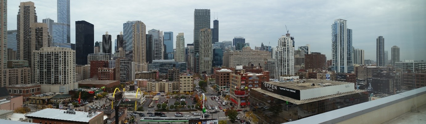
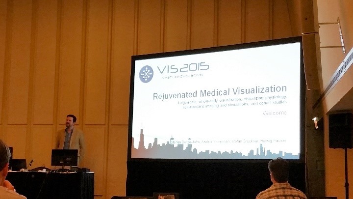
This year IEEE VIS (the conference formerly known as VisWeek) 2015 takes place in Chicago, also known as the “Windy City”. On the first day, I attended a very interesting tutorial about medical visualization entitled “Rejuvenated Medical Visualization”. This tutorial was opened by Steffen Oeltze-Jafra, the organizer of this event. He welcomed the audience and introduced the other speakers in the tutorial: Anders Ynnerman, Stefan Bruckner and Helwig Hauser.
Attending VIS 2015 in Chicago this year? Lucky you! We’ve compiled an overview of papers that sound relevant to our medical visualization interests and have some recommendations on what workshops, tutorials and sessions to attend to maximize your medvis experience. Can’t make it this year? No worries, there will be a full conference report afterwards.
If VA is not your jam, I recommend going to this tutorial: Rejuvenated Medical Visualization—LargeScale, Whole-Body Visualization, Visualizing Physiology, Non-standard imaging and Simulations, and Cohort Studies featuring Steffen Oeltze-Jafra, Anders Ynnerman, Stefan Bruckner and Helwig Hauser from 8:30am–12:10pm at the Monroe. Check the trailer:
In the afternoon you can then stay in the Monroe for the tutorial on Direct Volume Interaction for Visual Data Analysis with Alexander Wiebel, Tobias Isenberg, Stefan Bruckner and Timo Ropinski from 14:00-17:55. There’s a video too:
Monday:
If you’re interested in uncertainty visualization, there is a full day workshop (8:30am-5:55pm) in the Empire room: Visualization for Decision Making Under Uncertainty featuring Kristin Potter, Ruediger Westermann, Christoph Heinzl, Mike Kirby, Ross Whitaker, Eduard Groller, Torsten
Möller and Stefan Bruckner.
If you’re up for some Paraview in the afternoon there is a tutorial (2:00pm-5:55pm) in the Adams room, appropriately entitled The ParaView Tutorial
In the evening (7:00pm-9:00pm) there is a Practicioner event in the Empire, featuring a talk on “Visual Exploration in Surgery Monitoring for Coronary Vessels”:
Tuesday:
The first SciVis session in the State “SciVis Intro + Biomedical and Molecular Visualization (I)” (10:30am-12:10pm) is a definite must-see:
Wednesday morning you have a difficult choice to make ^^, both the SciVis and VAST sessions look interesting:
SciVis in the State: Tasks and Applications (8:30am-10:10am):
A Classification of User Tasks in Visual Analysis of Volume Data (C)
Authors: Bireswar Laha, Doug Bowman, David Laidlaw, John Socha Video Preview
Using Maximum Topology Matching to Explore Differences in Species Distribution Models (C)
Authors: Jorge Poco, Harish Doraiswamy, Marian Talbert, Jeffrey Morisette, Claudio Silva Video Preview
Visual Verification of Space Weather Ensemble Simulations (C)
Authors: Alexander Bock, Asher Pembroke, M. Leila Mays, Lutz Rastaetter, Anders Ynnerman, Timo Ropinski Video Preview
A Visual Voting Framework for Weather Forecast Calibration (C)
Authors: Hongsen Liao, Yingcai Wu, Li Chen, Thomas M. Hamill, Yunhai Wang, Kan Dai, Hui Zhang, Wei Chen Video Preview
Real-time Uncertainty Visualization for B-Mode Ultrasound (C)
Authors: Christian Schulte zu Berge, Denis Declara, Christoph Hennersperger, Maximilian Baust, Nassir Navab
At the same time, the VAST session in the Red room, Visual Analytics in Medicine and Healthcare, also looks tempting (8:30am-10:10am):
Integrating Predictive Analytics into a Spatio-Temporal Epidemic Simulation (C)
Authors: Chris Bryan, Xue Wu, Susan Mniszewski, Kwan-Liu Ma Video Preview
The SciVis sessions after the morning session are
Feature Extraction and Flows (10:30am-12:10pm) and Maps, Geometry and Terrain (4:15pm-5:55pm), neither of which seem to contain any medvis.
Thursday:
Thursday morning starts with another interesting SciVis session in the State room Interfaces, Languages and Systems (8:30am-10:10am):
The CG&A session at the Empire deals with Application-Tailored Visualizations (2:00pm-3:40pm) and features among other topics, this medvis-related paper:
Interactive Visual Analysis of Heterogeneous Cohort Study Data
Authors: Paolo Angelelli, Steffen Oeltze, Judit Haász, Cagatay Turkay, Erlend Hodneland, Arvid Lundervold, Astri J. Lundervold, Bernhard Preim, Helwig Hauser
Friday:
Friday morning starts with part two our favorite session topic Biomedical and Molecular Visualization (II) in the State (8:30am-10:10am):
Exploration of the Brain’s White Matter Structure through Visual Abstraction and Multi-Scale Local Fiber Tract Contraction (T)
Authors: Maarten H. Everts, Henk Bekker, Jos B.T.M. Roerdink, Tobias Isenberg
Medvis.org-contributor Kai Lawonn will be representing team medvis.org at the event, so be sure to go say hi to him if you see him (you might get one of the extremely-limited-edition medvis.org business cards ;)). Besides representing, he’s also presenting his work Tuesday morning in the ‘SciVis Intro + Biomedical and Molecular Visualization
(I)’-session, so don’t miss that one either ^^. I would like to wish all attendees a great time at VIS! Good luck and have fun! For those left behind like me, I can only wish Scivis does a little tweeting for a change 😉
Update: The slides of all talks are now available here!
MICCAI 2015! This year, MICCAI (International Conference on Medical Image Computing and Computer Assisted Intervention) takes place in Munich almost during the Oktoberfest. Sauerkraut, Weißwurst, Beer, and Science. What a great combination! The first day, MICCAI started with a few satellite events. Among these events, for us most relevant: the Tutorial on Advanced Medical Visualization. Most of the big shots in the medical visualization area contributed to this event and talked about ongoing research and the current state of the art in medical visualization. Anna Vilanova and Bernhard Preim were the hosts of this tutorial and introduced it.
Recently I had the pleasure of attending the Eurographics Workshop on Visual Computing for Biology and Medicine (VCBM) 2015 conference for the third and potentially, but not hopefully, last time. This year it was held in Chester (UK) at The Riverside Innovation Centre at the University of, you guessed it, Chester! In this conference report I will summarize some personal highlights. Repeating last year’s tradition, I again tweeted a picture for almost every talk. I still don’t think Twitter is really gaining traction among the scivis community, and I wonder what it would take to change it (or if it even needs to change ^^). As every year, given the theme of the conference almost every talk is relevant to our medical visualization interests, but I would like to briefly summarize only a couple of them here. Check the full list of papers and posters presented here if this is not enough to satiate your VCBM-craving-needs. Continue reading →
As we mentioned previously, you’ll be able to interact personally with many of Medical Visualization’s most handsome and knowledgable professors, and even some from medical imaging: Stefan Bruckner, Katja Bühler, Thomas Deserno, Eduard Gröller, Markus Hadwiger, Bernhard Kainz, Wiro Niessen, Bernhard Preim, Timo Ropinski, Thomas Schultz, Anna Vilanova, Rüdiger Westermann and Anders Ynnerman.
Sharing of this post with interested friends and colleagues would be most appreciated! (see the sharing icons right below this)
The “Call for Papers and Posters” for VCBM (AKA Eurographics Workshop on Visual Computing for Biology and Medicine) 2015 was released a while ago. As you probably already know, VCBM is an excellent venue for medical visualization work and this year it will be held in Chester, UK.
The deadline for full paper submission is June 21st and the posters need to be submitted by August 7th. Don’t miss this opportunity to present your work at this excellent location and please take a look at the website for more details.
(Cross-posted from medvisbook.com: The go-to resource for all things related to the book ‘Visual Computing for Medicine – Second Edition’. Original post written by Charl Botha.)
Bernhard Preim (University of Magdeburg, DE; author of and driving force behind the MedVis book) and Anna Vilanova (Delft University of Technology, NL) are organizing a tutorial on Advanced Medical Visualization at MICCAI 2015, one of the most important technical medical imaging conferences in the world today.
Judging by the list of topics and especially the list of speakers, I expect that this is going to be a great tutorial. If you’re going to MICCAI 2015, don’t miss this!
In the spirit of better late than never, the Eurographics Workshop on Visual Computing for Biology and Medicine (VCBM) 2014 conference report, summarizing some personal highlights. At VCBM 2014, we tweeted a picture for every talk and then some, which was arguably a bit much, but still a lot of fun. The VCBM organization also posted a great recap using Storify at their website. Given the theme of the conference, almost every talk is relevant to our medical visualization interests, but I would like to briefly summarize only a couple of them here. The benefit of delaying so long in writing this is that there are a lot of videos online by now. I will try to let the videos speak a 1000 words where available instead of getting too verbose. Onwards to the highlights!
The venue was really amazing, VCBM was held in the Universitätscampus
Altes AKH, but not just in any old lecture hall, it was the former anatomical theatre of the AKH and still has the original marble slab that was used as a dissection table:
Hörsaal D at the AKH
A day before the start of VCBM itself, the VCBM fachgruppe (working group) had a meeting with six interesting talks. This was followed by a social event, a guided tour of the Narrenturm. Built in 1874 to treat mental patients, it now serves as a museum for the Pathologic-Anatomical Collection. The Narrenturm features a huge collection of moulages. These are wax models of diseases made based on real patients and used in medical education, which is cool and slightly creepy at the same time. This tour was followed up by a delicious dinner at Unibräu for those who didn’t lose their appetite after what they had seen during the tour.
On Thursday, VCBM itself kicked off with an opening by Katja Bühler. After this we enjoyed a keynote by Anna Vilanova on the future of medical visualization. Anna presented medical visualization as a field that is between fields: computer graphics and medical imaging. She talked us through the past, present and future of medvis and going from facilitating analysis of the known to unraveling the unknown using visualization. A memorable quote from her talk:
“If the brain were so simple that we could understand it, we would be so simple we couldn’t” – Lyall Watson
Thursday featured four interesting sessions on Multivariate Data Analysis, Segmentation and Uncertainty, Microscopy and Visual Analytics for Biology:
Multivariate Data Analysis:
Benjamin Köhler presented interesting work on “Robust Cardiac Function Assessment in 4D PC-MRI Data” [1]. Four-dimensional phase-contrast magnetic resonance imaging (4D PC-MRI) is a relatively new modality that can acquire non-invasive, time-resolved, three-dimensional blood flow information. Benjamin proposed robust quantification techniques to assess cardiac function in this data.
I am obviously biased because she is from my group ;), but Renata Raidou presented her work entitled “The iCoCooN: Integration of Cobweb Charts with Parallel Coordinates for Visual Analysis of DCE-MRI Modeling Variations” [2]. She proposed a visualization application for the exploration and analysis of model-induced
variations in pharmacokinetic parameters. For this application, she created a visual representation, the Cocoon, by integrating perpendicularly Parallel Coordinate Plots (PCPs) with Cobweb Charts (CCs). A demo video is available here.
Segmentation and Uncertainty:
Peter Faltin presented his work on “Extracting and Visualizing Uncertainties in Segmentations from 3D Medical Data” [3]. He introduces a new processing chain comprising a series of carefully selected and well-matched steps to
determine and visualize a segmentation boundary. Additionally, a novel visualization method was presented, specifically designed to simultaneously provide information about 3D morphology, confidence and possible errors.
Microscopy:
Steffen Oeltze-Jafra delivered an excellent talk on “Interactive Labeling of Toponome Data” [4]. In this work, they present an approach for the in-place annotation of multi-channel microscopy data in 2D views, combining dynamic excentric labeling and static necklace maps:
Visual Analytics for Biology:
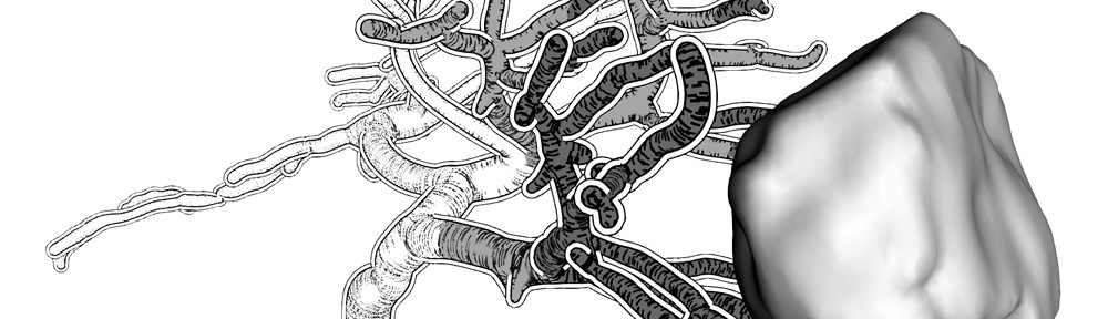
I really enjoyed the talk by Nicolas Swoboda on “Visual and Quantitative Analysis of Higher Order Arborization Overlaps for Neural Circuit Research” [5]. The overlaps they are reffering to, consist of two or more neurons and indicate a potential anatomical connection. They present a novel tool for potential connectivity exploration by providing for the first time the possibility to compute and visualize higher order arborization overlaps on the fly (for fruit fly brains, well played!) and to interactively explore this information in its spatial
anatomical context and on a quantitative level. Slides of the talk are available here and this is the accompanying video:
In the evening we hiked up through the vineyards of Vienna to the main social event: dinner at the Waldgrill Cobenzl. The view on the vineyards and Vienna itself was really stunning. We enjoyed a delicious buffet dinner accompanied by Sturm. Sturm is grape juice that has just started fermenting and is only available for a limited time every year, so we were lucky VCBM was held in Vienna exactly during Sturm time. After dinner the winners of the Karl-Heinz Höhne Award for Medical Visualization were announced:
I would love to tell you who the winner’s were, but the official announcement has not been made yet ;), so I don’t dare… Congratulations to the award winnners nonetheless, you know who you are ^^.
On the second and last day of VCBM there were sessions on Volume Visualization, Image Registration and Data Reconstruction for Medical Interventions, Visual Explanations and Display Techniques as a keynote by Nigel John entitled ‘Visual Computing in Healthcare – from the Research Lab into the Hospital”. In the keynote he presented several case studies and discussed some of the challenges
involved in deploying visual computing solutions in a hospital setting.
Stefan Lindholm gave an interesting talk entitled “Towards Clinical Deployment of Automated Anatomical Regions-Of-Interest” [7]. In his work he proposes an Automatic and Anatomical ROI (AA-ROI) approach based on the combination of automatic image registration (AIR) and distance-based Transfer Functions (DBTFs), designed for automatic selection of complex anatomical shapes without relying on excessive amounts of interaction:
Towards Clinical Deployment of Automated Anatomical Regions-Of-Interest
Image Registration and Data Reconstruction for Medical Interventions:
At the risk of tooting my own horn, I gave a talk on “RegistrationShop: An Interactive 3D Medical Volume Registration System” [8], developed to improve and simplify the process of volume registration with 3D visualizations and
simple interactive tools. The source code is available here, talk slides are here, and this is a demo video:
The honorable mentions can be found here. Our congratlations to the authors! Ivan Viola closed the conference and announced the location for next year (this year by now ^^): VCBM 2015 will be held in Bangor (UK) and will from now on be an annual workshop instead of bi-annual (once every two years, not the twice every year-type). To conclude this summary, I’d really like to thank the organizers of this excellent workshop. Interesting talks, a beautiful location, good food, great people once again!
References:
[1]: Robust Cardiac Function Assessment in 4D PC-MRI Data. Köhler, Benjamin; Preim, Uta; Gutberlet, Matthias; Fischbach, Katharina; Preim, Bernhard
[2]: The iCoCooN: Integration of Cobweb Charts with Parallel Coordinates for Visual Analysis of DCE-MRI Modeling Variations. Raidou, Renata; Breeuwer, Marcel; Vilanova, Anna
[3]: Extracting and Visualizing Uncertainties in Segmentations from 3D Medical Data. Faltin, Peter; Chaisaowong, Kraisorn; Kraus, Thomas; Merhof, Dorit
[4]: Interactive Labeling of Toponome Data. Oeltze-Jafra, Steffen; Pieper, Franz; Hillert, Reyk; Preim, Bernhard; Schubert, Walter
[5]: Visual and Quantitative Analysis of Higher Order Arborization Overlaps for Neural Circuit Research. Swoboda, Nicolas; Moosburner, Judith; Bruckner, Stefan; Yu, Jai Y.; Dickson, Barry J.; Bühler, Katja
[6]: Visibility-Driven Processing of Streaming Volume Data. Solteszova, Veronika; Birkeland, Åsmund; Viola, Ivan; Bruckner, Stefan
[7]: Towards Clinical Deployment of Automated Anatomical Regions-Of-Interest. Lindholm, Stefan; Forsberg, Daniel; Ynnerman, Anders; Knutsson, Hans; Andersson, Mats; Lundström, Claes
[8]: RegistrationShop: An Interactive 3D Medical Volume Registration System. Smit, Noeska; Klein Haneveld, Berend; Staring, Marius; Eisemann, Elmar; Botha, Charl; Vilanova, Anna
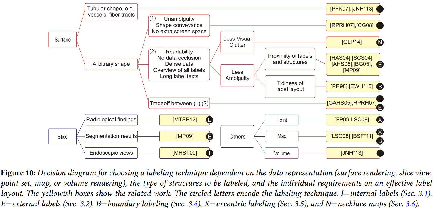
[9]: Survey of Labeling Techniques in Medical Visualizations. Oeltze-Jafra, Steffen; Preim, Bernhard