The VCBM (AKA Eurographics Workshop on Visual Computing for Biology and Medicine) submission deadlines are approaching! The deadline for full paper submission is June 29 and the posters need to be submitted by August 10. So if you’d like to present your work at this excellent location, please take a look at this page.
Open Positions at KAUST – Geometric Modeling and Scientific Visualization Center (Saudi Arabia)
I just returned from Eurovis 2012 and while a more detailed write-up of that will be posted soon, I wanted to let you know about several open positions over at KAUST in Saudi Arabia in the meantime.

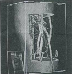
An example of the research done at GMSV 'Fused Multi-Volume DVR using Binary Space Partitioning'
The newly formed Geometric Modeling and Scientific Visualization Center (GMSV) is looking for 5-10 post-docs, MS and PhD students as well as short term visitors (2-3 months). A overview of their scivis work so far can be seen here. Contact them by sending an email to gmsvfaculty(a)kaust.edu.sa.
Two Medical Visualisation Ph.D. Vacancies at VRVis in Vienna (Austria)
Katja Bühler’s group at VRVis in Vienna currently has two medical visualisation Ph.D. vacancies. They are looking for two highly motivated young scientists preferably from European Countries interested in medical visualization and in the development of cutting edge solutions for radiotherapy planning.
More details and the description for these jobs can be found here
The lucky person that gets the first position can start right away and the second position will be available from August onwards.
Groundbreaking volume rendering papers by Professor Karl Heinz Höhne: a medvis.org exclusive!
As medvis professionals, we are accustomed to volume rendering as an every-day tool for exploration of medical data. The two technical papers below represent one of the first milestones in rendering volumetric data. We are extremely happy and excited that Professor Karl Heinz Höhne allowed us to post these papers on our blog. Published in 1986 and 1988, these papers cannot be found online anywhere else and proposed groundbreaking volume rendering techniques.
The first paper entitled ‘Shading 3D-images from CT Using Gray-Level gradients’ by Karl Heinz Höhne and Ralph Bernstein was published in 1986 in the IEEE Transactions on Medical Imaging. This paper describes a shading method based on the partial volume effect using the gray-level gradients along a surface reconstruction of CT images. What sets this method apart from current gradient vector computations is that the gray-values are sampled in screen space and not in voxel space. As a result, surfaces perpendicular to the viewer are brighter than surfaces oriented away from the viewer. In essence, this shading method simulates a headlight configuration.
- Shading 3D-images from CT Using Gray-Level gradients – hohne86
- Shading 3D-images from CT Using Gray-Level gradients correction – hohne86
Using the software and hardware that was available at the time, these papers represent many contributions to the field, for instance selective volume clipping and multimodal visualization. With their work, they created a solid foundation for state-of-the-art (medical) volume rendering techniques. The two papers usually credited as first presenting the ideas behind raycasting, both published in 1988, are:
- R. A. Drebin, L. Carpenter, and P. Hanrahan, “Volume rendering,” SIGGRAPH Comput. Graph., vol. 22, no. 4, pp. 65–74, Aug. 1988.
- M. Levoy, “Display of surfaces from volume data,” IEEE Computer Graphics and Applications, vol. 8, no. 3, pp. 29-37, May 1988.
Visualisation professorship at the University of Bergen (Norway)
The Department of Informatics at the University of Bergen has the opportunity to hire a new professor (a full, i.e., tenured professor) into the visualization research group with the overall goal to achieve a good complementary fit with the existing research/teaching profile in visualization.
The position has been announced via the jobbnorge-portal, together with a short description of the environment and the plans with regard to the new professor (see also the attached PDF). The application deadline is set to Dec. 22nd, 2011.
Visualisation Ph.D. vacancy at the University of Bergen (Norway)
The Department of Informatics at the University of Bergen is hiring a PhD candidate in informatics (but can be visualisation, according to Helwig Hauser). Don’t miss out on this amazing opportunity to become a legendary Visualisation Ninja at one of the best visualization places in the world.
More details and the job description can be found here. The deadline for applying is the 15th of December.
Report on the Medical Visualization session at VisWeek 2011
(We are happy and grateful that Dr. Steffen Oeltze from the University of Magdeburg Visualization Group could write this short report on the medical visualization session and other medvis-related papers at IEEE VisWeek 2011.)
This year, the IEEE VisWeek has been completed by an excellent session on medical visualization hosting five contributions from three European countries. Roy van Pelt gave a compelling talk on the exploration of cardiovascular 4D MRI blood-flow using stylistic visualizations. His comic-inspired illustrative glyphs coupled with timelines outperform traditional particle renderings. Interactive virtual probing of the flow
avoids a tedious segmentation process in qualitative inspection.
Rostislav Khlebnikov presented a new approach to tumor accessibility planning. It exploits a well-known natural phenomenon related to light scattering at dust particles which is also called crepuscular rays. In the generated 2D/3D images, light beams in different colors that shine through the skin indicate the access paths and their associated risk.
Christian Dick presented new visualization techniques for conveying distances in interactive 3D implant planning. The design of very intuitive distance glyphs and colored slice sets was completed by a carefully accomplished, convincing user study.
Rocco Gasteiger introduced the FlowLens for focus+context visualization of blood flow in cerebral aneurysms. It supports an exploration of certain hemodynamic attributes in the lens region within the context of other attributes thereby avoiding the cognitive effort involved in mental superimposition of side-by-side visualizations. Please watch the supplemental video:
The session was completed by the interesting talk of Artem Amirkhanov on the reduction of metal artifacts in industrial 3D X-ray CT images. He presented a projection-space pipeline in which metal is separated from the other materials before projection and then fused again with the initial reconstruction after projection.
Other talks not being part of the session but also related to medical visualization were given by Claes Lundström on the application of a multi-touch table system to orthopedic surgery planning, Christian Rieder on real-time approximation of the ablation zone for radiofrequency ablation (see the very nice video), Joseph Marino on context preserving maps of tubular structures, e.g., the colon, and Paolo Angelelli on straightening aortic blood flow for side-by-side visualization.
Exposure Render 1.1 now available!
Exposure Render is the open source GPU volume rendering implementation by Thomas Kroes. What makes this different from all other DVR implementations is the fact that it supports physically-based lighting at interactive speeds. So what does all of that mean? It means that you can make incredibly pretty interactive volume renderings on your own PC. See this youtube movie for example:
… or the rendering below (one of my favourites):
Read more about Exposure Render by going to its TU Delft Graphics website, or to the Google Code project page where you can download and play with the software!
P.S. Don’t forget to cite the paper when it gets published.
MedVis Ph.D. vacancy at the TU Delft
The Medical Visualisation group at the TU Delft currently has a fully funded 4-year Ph.D. vacancy. The successful candidate will work together with another Ph.D. student from the LUMC Departments of Anatomy and Surgery on building the Virtual Surgical Pelvis, a next generation surgical model, in silico, of the human pelvis, based on extremely high resolution histological sections. For more details on the project and the vacancy, see this webpage.
The deadline for applying is May 15, so you better hurry up!
Virtual Autopsy table now available commercially
Engadget reports that the virtual autopsy table we posted about in October of last year, is now commercially available from Sectra as the Visual Imaging Table. Get yours now while stocks last.
Chalk another one up for MedVis in the mainstream!


