There is an open position for a PhD at Aviz-Inria in France. They are looking for someone to work on a project on the interesting topic of Structural and Functional Visualization of Brain Connectivity. The project combines the best of both worlds with a mix scivis and infovis approaches. Does this sound like your type of project? If so, keep in mind the application deadline is August 1, 2015 and start working on your application. More information on the project and the position can be found in this pdf.
The Eurographics Association organizes a biannual competition to acknowledge the contribution that computer graphics is playing in the medical field, and to encourage further development. In 2010 the Eurographics Medical Prize was renamed to the Dirk Bartz Prize for Visual Computing in Medicine to honor Dirk Bartz who passed away far too early in March the same year. Dirk was a highly recognized and enthusiastic scientist, teacher and promoter of Visual Computing in Medicine, an active member of the Eurographics Association, and Chair of the EG Medical Prizes 2007 and 2009.
At this year’s Eurographics conference, the three winners of the Dirk Bartz Prize for Visual Computing in Medicine 2015 were announced. The three winning contributions were selected by an international program committee that was chaired by Hans Christian Hege and Timo Ropinski. Based on the reviews of this committee consisting of computer scientists and medical experts, the following ranking was established:
3rd Prize: Multi-Touch Table System for Medical Visualization by Anders Ynnerman, Thomas Rydell, Anders Persson, Aron Ernvik, Camilla Forsell, Patric Ljung, and Claes Lundström
We would like to congratulate all winning teams!
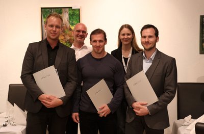
Hugo Talbot (left) receives the first prize from the EG Medical Prize Chairs (Timo Ropinski centre, Hans Christian Hege right) at this year’s EG Medical Prize ceremony.
Every two years the VCBM group awards the Karl-Heinz Höhne Award for Medical Visualization (medvis-award for short) to a young scientist in the MedVis field. Candidates for this award focus their innovative research on visualizations clearly related to medical questions.
The official website has not been updated yet, but we have received a tip from Frank Heckel that there has been a press-release announcing the winners (in German). Last year at VCBM, three contestants got awarded for their medical visualization work:
In third place: Cees-Willem Hofstede (Delft University of Technology) for his work on the Online Anatomical Human: an online anatomy browser, which allows combined display of medical images and reconstructed 3D models of various anatomical structures.
In second place: Frank Heckel (Fraunhofer MEVIS) for his work on interactive correction of segmentation results, such as delineated tumors in CT-scans.
In first place: Benjamin Köhler (Universität Magdeburg) for his work on the quantification and visual representation of blood flow data in cardiovascular disease.
Karl-Heinz-Höhne Award (MedVis- Award) winners: from left to right: Cees-Willem Hofstede, Benjamin Köhler and Frank Heckel in the front row. Dr. Stefan Zachow and Dr. Dorit Merhof in the back row.
We would like to congratulate the winners with their well-deserved awards!
(Cross-posted from medvisbook.com: The go-to resource for all things related to the book ‘Visual Computing for Medicine – Second Edition’. Original post written by Charl Botha.)
Bernhard Preim (University of Magdeburg, DE; author of and driving force behind the MedVis book) and Anna Vilanova (Delft University of Technology, NL) are organizing a tutorial on Advanced Medical Visualization at MICCAI 2015, one of the most important technical medical imaging conferences in the world today.
Judging by the list of topics and especially the list of speakers, I expect that this is going to be a great tutorial. If you’re going to MICCAI 2015, don’t miss this!
In the spirit of better late than never, the Eurographics Workshop on Visual Computing for Biology and Medicine (VCBM) 2014 conference report, summarizing some personal highlights. At VCBM 2014, we tweeted a picture for every talk and then some, which was arguably a bit much, but still a lot of fun. The VCBM organization also posted a great recap using Storify at their website. Given the theme of the conference, almost every talk is relevant to our medical visualization interests, but I would like to briefly summarize only a couple of them here. The benefit of delaying so long in writing this is that there are a lot of videos online by now. I will try to let the videos speak a 1000 words where available instead of getting too verbose. Onwards to the highlights!
The venue was really amazing, VCBM was held in the Universitätscampus
Altes AKH, but not just in any old lecture hall, it was the former anatomical theatre of the AKH and still has the original marble slab that was used as a dissection table:
Hörsaal D at the AKH
A day before the start of VCBM itself, the VCBM fachgruppe (working group) had a meeting with six interesting talks. This was followed by a social event, a guided tour of the Narrenturm. Built in 1874 to treat mental patients, it now serves as a museum for the Pathologic-Anatomical Collection. The Narrenturm features a huge collection of moulages. These are wax models of diseases made based on real patients and used in medical education, which is cool and slightly creepy at the same time. This tour was followed up by a delicious dinner at Unibräu for those who didn’t lose their appetite after what they had seen during the tour.
On Thursday, VCBM itself kicked off with an opening by Katja Bühler. After this we enjoyed a keynote by Anna Vilanova on the future of medical visualization. Anna presented medical visualization as a field that is between fields: computer graphics and medical imaging. She talked us through the past, present and future of medvis and going from facilitating analysis of the known to unraveling the unknown using visualization. A memorable quote from her talk:
“If the brain were so simple that we could understand it, we would be so simple we couldn’t” – Lyall Watson
Thursday featured four interesting sessions on Multivariate Data Analysis, Segmentation and Uncertainty, Microscopy and Visual Analytics for Biology:
Multivariate Data Analysis:
Benjamin Köhler presented interesting work on “Robust Cardiac Function Assessment in 4D PC-MRI Data” [1]. Four-dimensional phase-contrast magnetic resonance imaging (4D PC-MRI) is a relatively new modality that can acquire non-invasive, time-resolved, three-dimensional blood flow information. Benjamin proposed robust quantification techniques to assess cardiac function in this data.
I am obviously biased because she is from my group ;), but Renata Raidou presented her work entitled “The iCoCooN: Integration of Cobweb Charts with Parallel Coordinates for Visual Analysis of DCE-MRI Modeling Variations” [2]. She proposed a visualization application for the exploration and analysis of model-induced
variations in pharmacokinetic parameters. For this application, she created a visual representation, the Cocoon, by integrating perpendicularly Parallel Coordinate Plots (PCPs) with Cobweb Charts (CCs). A demo video is available here.
Segmentation and Uncertainty:
Peter Faltin presented his work on “Extracting and Visualizing Uncertainties in Segmentations from 3D Medical Data” [3]. He introduces a new processing chain comprising a series of carefully selected and well-matched steps to
determine and visualize a segmentation boundary. Additionally, a novel visualization method was presented, specifically designed to simultaneously provide information about 3D morphology, confidence and possible errors.
Microscopy:
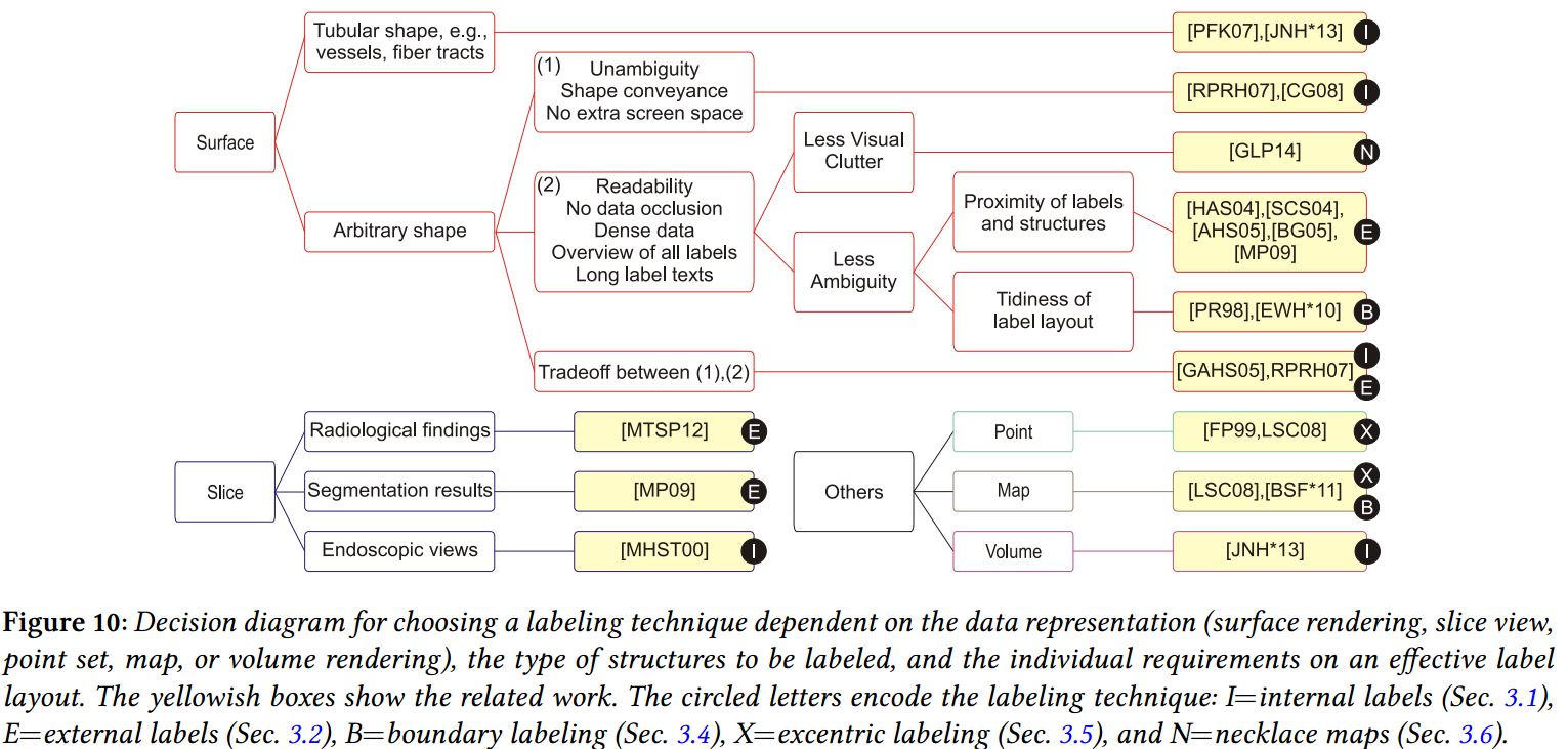
Steffen Oeltze-Jafra delivered an excellent talk on “Interactive Labeling of Toponome Data” [4]. In this work, they present an approach for the in-place annotation of multi-channel microscopy data in 2D views, combining dynamic excentric labeling and static necklace maps:
Visual Analytics for Biology:
I really enjoyed the talk by Nicolas Swoboda on “Visual and Quantitative Analysis of Higher Order Arborization Overlaps for Neural Circuit Research” [5]. The overlaps they are reffering to, consist of two or more neurons and indicate a potential anatomical connection. They present a novel tool for potential connectivity exploration by providing for the first time the possibility to compute and visualize higher order arborization overlaps on the fly (for fruit fly brains, well played!) and to interactively explore this information in its spatial
anatomical context and on a quantitative level. Slides of the talk are available here and this is the accompanying video:
In the evening we hiked up through the vineyards of Vienna to the main social event: dinner at the Waldgrill Cobenzl. The view on the vineyards and Vienna itself was really stunning. We enjoyed a delicious buffet dinner accompanied by Sturm. Sturm is grape juice that has just started fermenting and is only available for a limited time every year, so we were lucky VCBM was held in Vienna exactly during Sturm time. After dinner the winners of the Karl-Heinz Höhne Award for Medical Visualization were announced:
I would love to tell you who the winner’s were, but the official announcement has not been made yet ;), so I don’t dare… Congratulations to the award winnners nonetheless, you know who you are ^^.
On the second and last day of VCBM there were sessions on Volume Visualization, Image Registration and Data Reconstruction for Medical Interventions, Visual Explanations and Display Techniques as a keynote by Nigel John entitled ‘Visual Computing in Healthcare – from the Research Lab into the Hospital”. In the keynote he presented several case studies and discussed some of the challenges
involved in deploying visual computing solutions in a hospital setting.
Stefan Lindholm gave an interesting talk entitled “Towards Clinical Deployment of Automated Anatomical Regions-Of-Interest” [7]. In his work he proposes an Automatic and Anatomical ROI (AA-ROI) approach based on the combination of automatic image registration (AIR) and distance-based Transfer Functions (DBTFs), designed for automatic selection of complex anatomical shapes without relying on excessive amounts of interaction:
Towards Clinical Deployment of Automated Anatomical Regions-Of-Interest
Image Registration and Data Reconstruction for Medical Interventions:
At the risk of tooting my own horn, I gave a talk on “RegistrationShop: An Interactive 3D Medical Volume Registration System” [8], developed to improve and simplify the process of volume registration with 3D visualizations and
simple interactive tools. The source code is available here, talk slides are here, and this is a demo video:
The honorable mentions can be found here. Our congratlations to the authors! Ivan Viola closed the conference and announced the location for next year (this year by now ^^): VCBM 2015 will be held in Bangor (UK) and will from now on be an annual workshop instead of bi-annual (once every two years, not the twice every year-type). To conclude this summary, I’d really like to thank the organizers of this excellent workshop. Interesting talks, a beautiful location, good food, great people once again!
References:
[1]: Robust Cardiac Function Assessment in 4D PC-MRI Data. Köhler, Benjamin; Preim, Uta; Gutberlet, Matthias; Fischbach, Katharina; Preim, Bernhard
[2]: The iCoCooN: Integration of Cobweb Charts with Parallel Coordinates for Visual Analysis of DCE-MRI Modeling Variations. Raidou, Renata; Breeuwer, Marcel; Vilanova, Anna
[3]: Extracting and Visualizing Uncertainties in Segmentations from 3D Medical Data. Faltin, Peter; Chaisaowong, Kraisorn; Kraus, Thomas; Merhof, Dorit
[4]: Interactive Labeling of Toponome Data. Oeltze-Jafra, Steffen; Pieper, Franz; Hillert, Reyk; Preim, Bernhard; Schubert, Walter
[5]: Visual and Quantitative Analysis of Higher Order Arborization Overlaps for Neural Circuit Research. Swoboda, Nicolas; Moosburner, Judith; Bruckner, Stefan; Yu, Jai Y.; Dickson, Barry J.; Bühler, Katja
[6]: Visibility-Driven Processing of Streaming Volume Data. Solteszova, Veronika; Birkeland, Åsmund; Viola, Ivan; Bruckner, Stefan
[7]: Towards Clinical Deployment of Automated Anatomical Regions-Of-Interest. Lindholm, Stefan; Forsberg, Daniel; Ynnerman, Anders; Knutsson, Hans; Andersson, Mats; Lundström, Claes
[8]: RegistrationShop: An Interactive 3D Medical Volume Registration System. Smit, Noeska; Klein Haneveld, Berend; Staring, Marius; Eisemann, Elmar; Botha, Charl; Vilanova, Anna
[9]: Survey of Labeling Techniques in Medical Visualizations. Oeltze-Jafra, Steffen; Preim, Bernhard
We have received word from Bilal Alsallakh that the University of Bergen is looking for a PhD student in visualization. The exact topic of the PhD will be decided on with the advisor, but biomedical, visual analytics and visual computing are among the list of possible topics. More information about the position is available here. Alternatively you can skip to the full advertisement here and a factsheet here. The application deadline is the June 1, 2015, so do not delay and apply now if you are interested in this amazing opportunity.
I apologize for the delay in reporting this, but I heard about/attended three great medical visualization PhD defenses late 2014:
Dr. Mathias Goldau successfully defended his PhD at the University of Leipzig. His thesis is entitled ‘Multi-modal and Slice-based Visualizations of Diffusion Tractography Data’ and you may or may not already be familiar with his work on OpenWalnut, an open source tool for multi-modal medical and brain data visualization. The full text of the thesis can be downloaded here or take a look at his publications here.
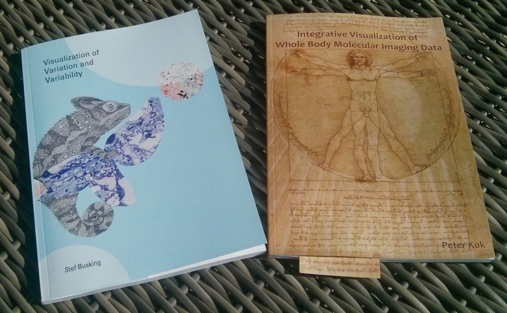
Dr. Peter Kok successfully defended his PhD at the Technical University of Delft. In his thesis, ‘Integrative Visualization of Whole Body Molecular Imaging Data‘, Dr. Kok present methods to map molecular imaging data to a common reference frame, to combine multiple modalities and to compare scans taken at different timepoints. The full text of the thesis is available here.
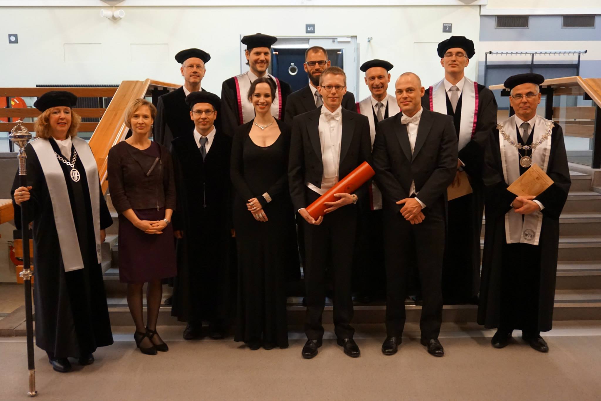
Dr. Kok with his paranymphs and the thesis committee members. tblr: Jos Roerdink, Elmar Eisemann, Charl Botha, Erik Jansen, Boudewijn Lelieveldt, beadle Rina Abbriata, Louise van der Weerd, Bernhard Preim, Noeska Smit, Peter Kok, Thomas Kroes and the head of the committee.
On the same day, Dr. Stef Busking successfully defended his PhD at the Technical University of Delft as well. His thesis, ‘Visualization of Variation and Variability‘ deals with comparative visualization as a means to analyze variation or variability based on two or more specific instances of the data. The full text of the thesis is available here.
This week the TU Delft in the Netherlands will host two medical visualization events in a single week:
First up, on Thursday December 11th, we will have a special medical visualization colloquium (open to the public). This colloquium will feature two invited speakers, Prof. Dr.-Ing. Bernhard Preim and Prof.dr. Jos Roerdink, presenting recent work on ‘Visual Analytics in Cohort Study Data’ and ‘Prediction of Neurodegenerative Diseases’. More information about the colloquium and the abstracts for the talks are available here.
Previously, we reported on two open positions in the context of the VAnPIRe, Visual Analysis in Population Imaging Research, project. The PhD position has since been filled, but the PostDoc is still open. More information about the position can be found here. The closing date is at the end of the month, so apply now if you’re interested!
We’re currently at VCBM in Vienna. For live updates, check out our Twitter coverage and be sure to check back for a full conference report in the near future!